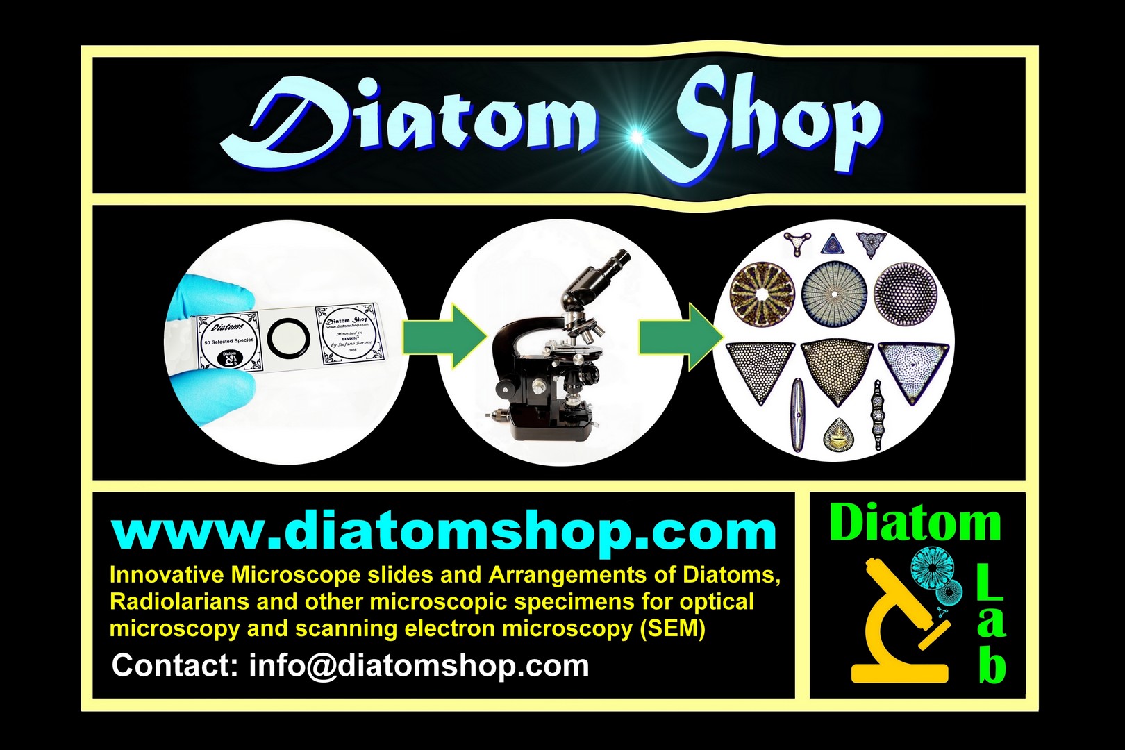
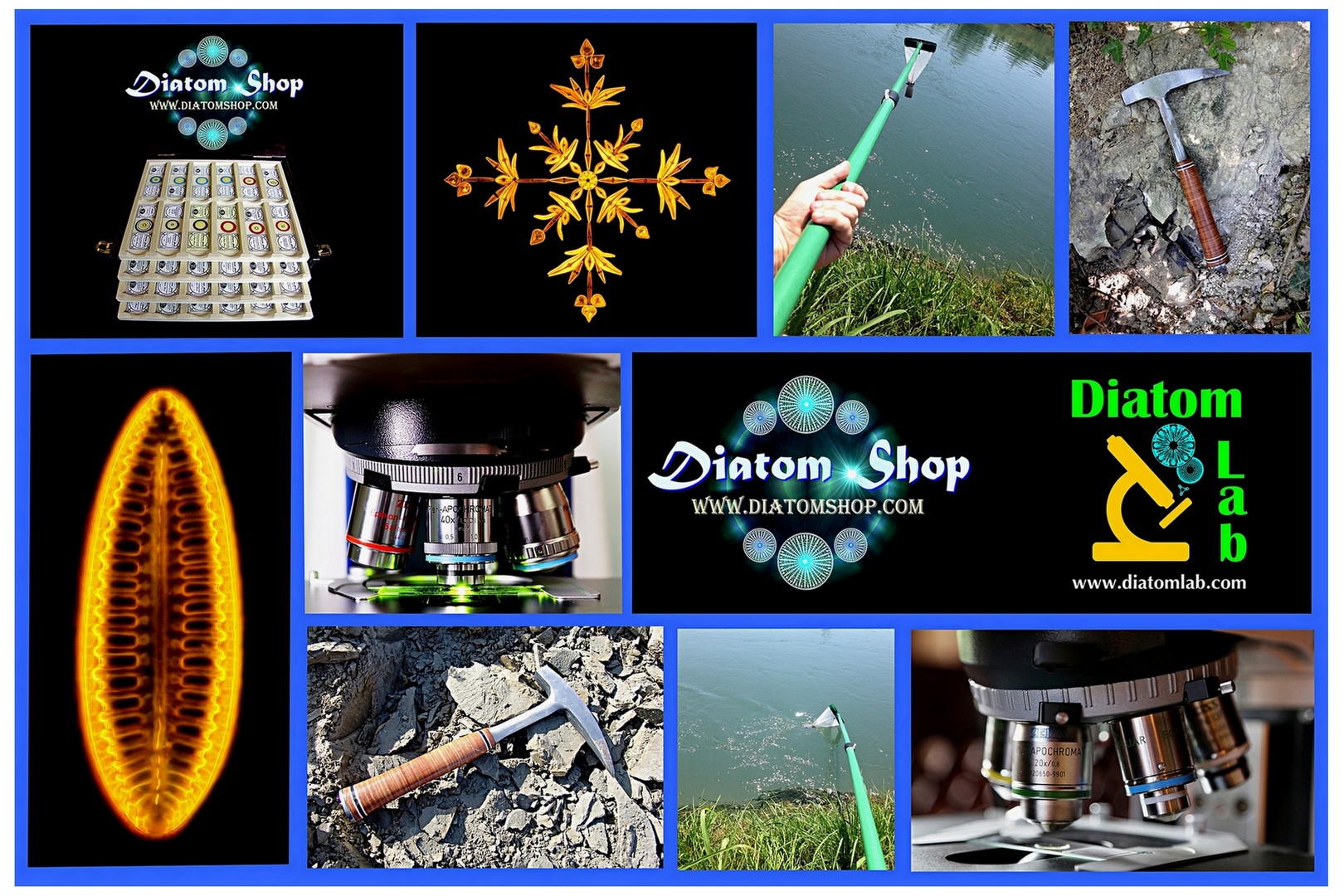
Welcome to Diatom Shop, the online store of Diatom Lab! Our preparations allows you to observe Diatoms and other microscopic objects in Very High Definition! Diatom Lab is the state-of-the-art scientific laboratory company founded in 2016 (VAT number IT 01635810193) that specializes in ground-breaking Micromanipulation services, Test slides for microscopy, Scientific photography / Microscopy imaging services.
Diatom Lab produces prepared microscope slides of the highest quality: our laboratory stands out for the use of unique cutting-edge materials, technologies and procedures. For further information please visit the pages "Innovative preparations" and "Diatom Lab. Clients and reputation".
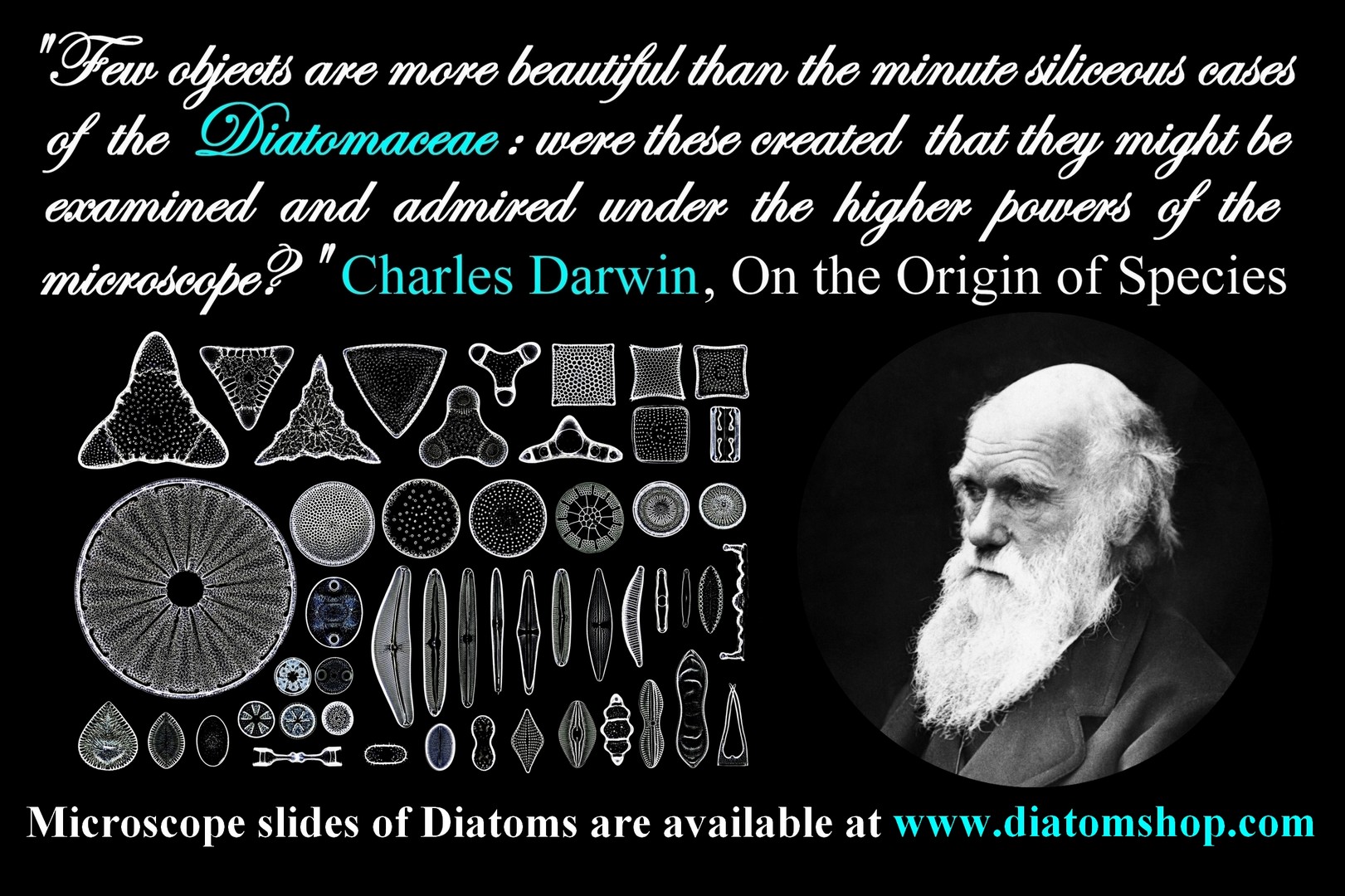
"Few objects are more beautiful than the minute siliceous cases of the Diatomaceæ: were these created that they might be examined and admired under the higher powers of the microscope?” (Charles Darwin, On the Origin of Species). Interesting facts about Diatoms: they are unicellular organisms, specifically microalgae, and live in waterways, oceans, and soils; they produce approximately 20 to 50 percent of the oxygen on Earth; they are primary producers in the food chain and constitute nearly half of the organic material found in the oceans; since the discovery of the microscope, architects and artists have been inspired by Diatom forms.
We safely ship Worldwide: all parcels are fully tracked, so that you can check the progress of your items through to delivery! We have customers in many countries including the UK, USA, Europe, Asia, Africa and Oceania, and look forward to working with you! Combined shipping: worldwide shipping by registered international parcel is always 11,90 euros, no matter how many items are in the cart! Payments are accepted with PayPal (no PayPal account needed, just a credit card!) or bank transfer. Diatom Lab works with honesty, accuracy and transparency: you'll always receive a proper invoice with every purchase.
If If you
are an institution or company and need an official quote in PDF format, please contact us and provide all the necessary data.
Click on the hamburger button at the top right to open the Menu.
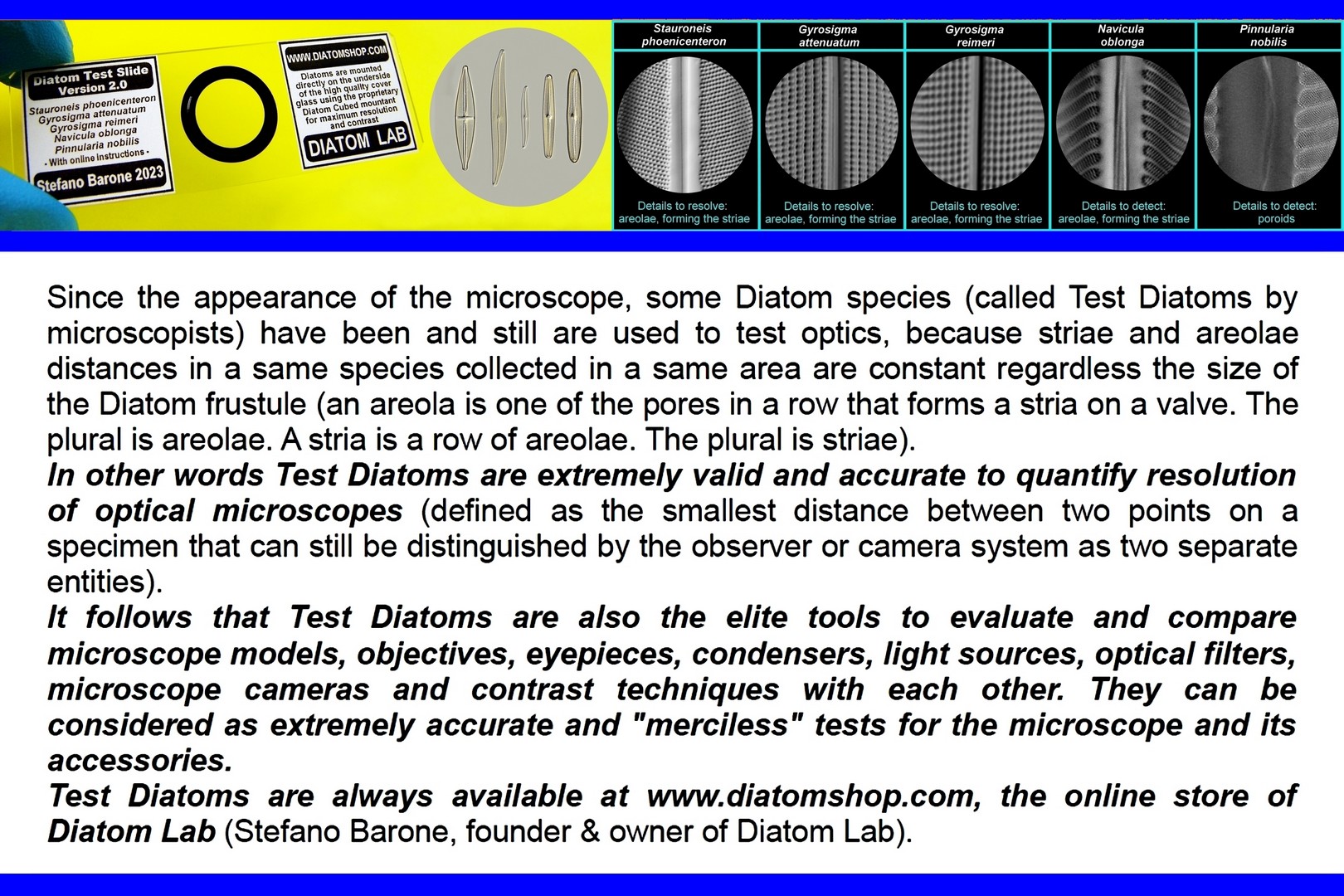
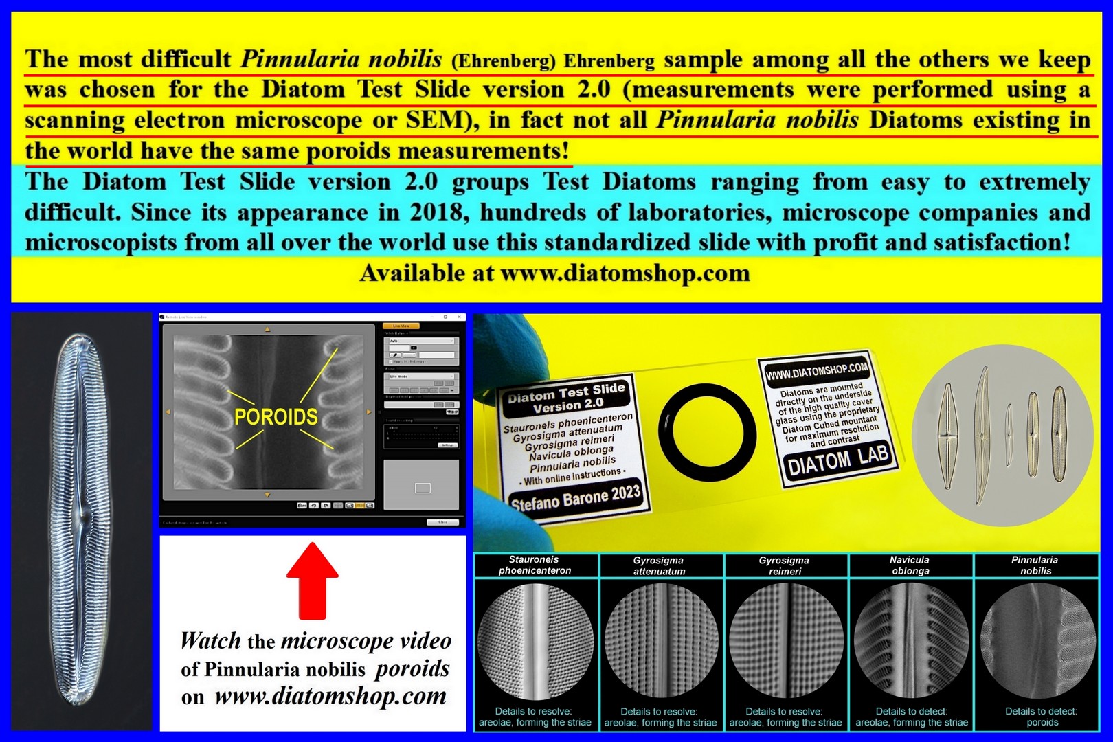
DIATOM TEST SLIDES FOR OPTICAL MICROSCOPES (MICROSCOPE SLIDES OF SELECTED, MICROMANIPULATED TEST DIATOMS). TEST YOUR MICROSCOPE! Venture into the Micro and Nano structures! DIATOM TEST SLIDES ARE EXTREMELY USEFUL TO: A) evaluate and compare microscope models, objectives, eyepieces, condensers, light sources, optical filters, microscope cameras and microscope illumination techniques such as bright field, oblique illumination, differential interference contrast (DIC), phase contrast, dark field, polarized light, Rheinberg illumination, and Hoffman modulation contrast! B) quantify resolution in optical microscopes (defined as the smallest distance between two points on a specimen that can still be distinguished by the observer or camera system as two separate entities); C) practice using your microscopes at their highest levels! D) examine the variations in contrast and resolution by regulating the condenser aperture diaphragm; E) understand the importance of correction collar for minimazing spherical aberration
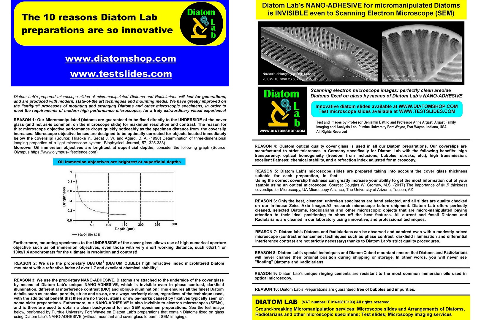
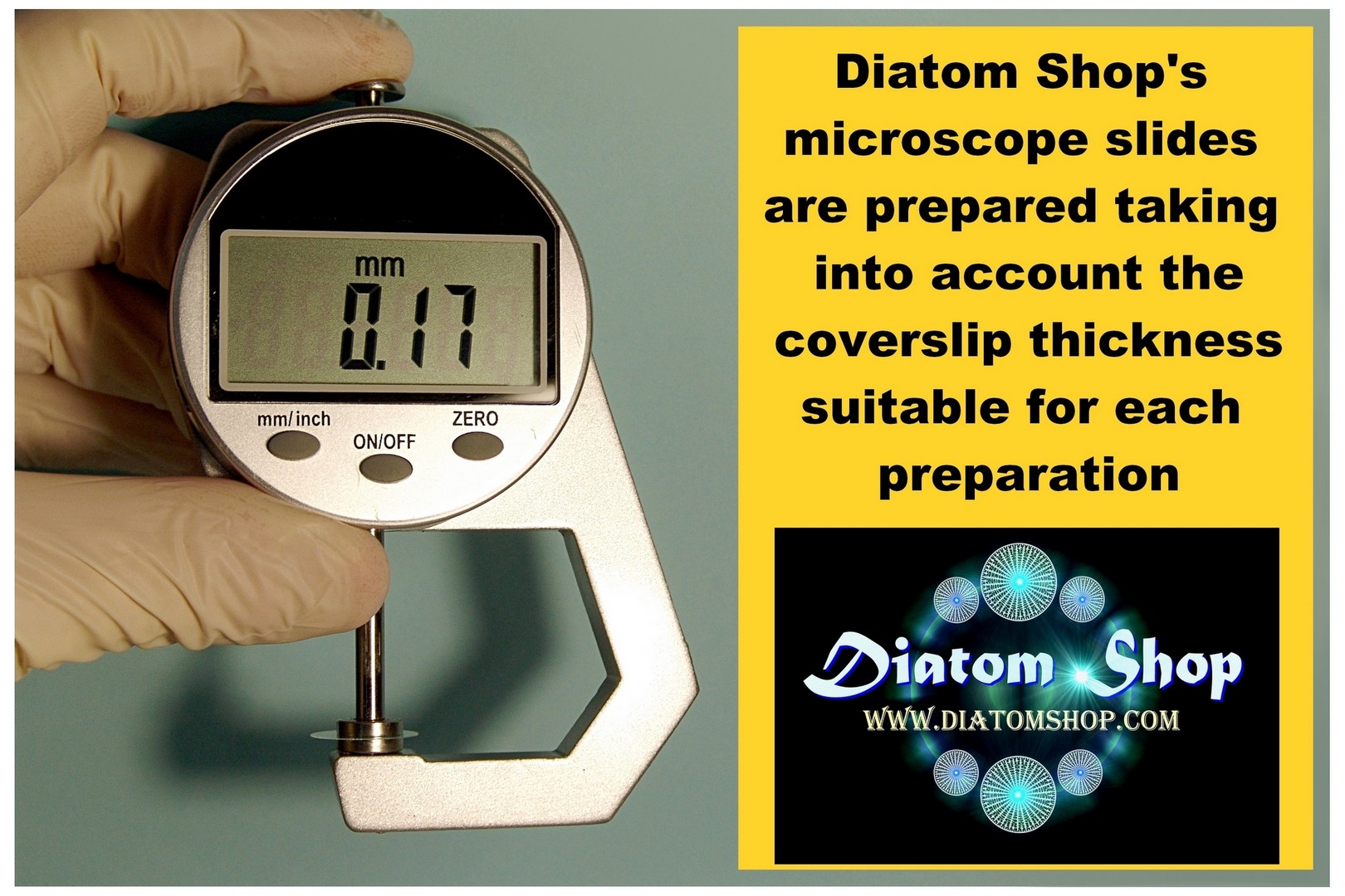
All micromanipulated Diatoms are guaranteed to be fixed directly to the UNDERSIDE of the custom, optical quality cover glass (and not, as is common, on the microscope slide) for maximum resolution and contrast! (The reason for this: microscope objective performance drops quickly and noticeably as the specimen distance from the cover glass increases. Microscope objective lenses are designed to be optimally corrected for objects located immediately below the coverslip!)
- Barone, S. 2023. Diatoms: the best microscopic objects to check, set and compare optical microscopes and contrast techniques, Microscopy and Analysis, 65: 13-18 (ISSN 2049-4424) link
- Barone S., 2022. Diatom Lab Microscope Test Slide in commemoration of Edmund J. Spitta, Micscape Magazine, 321 (ISSN 1365 - 070x) link
David Walker is the editor of Micscape Magazine:
- Walker D., 2023. Exploring the Diatom Lab 'Microscope Test Slide in Commemoration of Edmund J. Spitta' with Near UV . Micscape Magazine, 328 (ISSN 1365 - 070x) link
- Walker D., 2024. Exploring the Diatom Lab test slide Pinnularia dactylus var. dariana (A. Schmidt) Cleve 1895 at ca. 400 nm, Micscape Magazine, 339 (ISSN 1365 - 070x) link "Some time ago I treated myself to three single species diatom test slides prepared by Stefano Barone of Diatom Lab but it is only recently that I have started to explore them. From previous slides purchased I knew they were prepared to the very highest standards with the diatom touching the under side of the high spec. coverslip and use a proprietary Diatom Cubed high RI 1.7 mountant. A variety of test slides can be bought on the dedicated www.diatomshop.com website";
- Walker D., 2021. Note on 'diatom dotting' at low magnifications. Resolving Stauroneis phoenicenteron, Micscape Magazine, 306 (ISSN 1365 – 070x) “Acknowledgement: The author used the invaluable 'Test Slide version 2.0' (Diatom Cubed mountant) supplied by Stefano Barone of Diatom Lab"
- Bulletin of the Quekett Microscopical Club, Nov 2019, No.77, ISSN 1350-9128, pp 22-25: the article recognizes Diatom Lab preparations as expertly-prepared and three microscope images of our slides have been published, such as a detail of the Diatom Test Slide version 2.0 with the comment “image chosen by editor to show the incredible resolution of this excellent slide”
Diatom Test Slide version 2.0 Click on the PDF button to download the free instructions (Updated edition)  | Microscope Test Slide in commemoration of Edmund J. Spitta. Click on the PDF button to download the free instructions   | Free article: Barone, S. 2023. Diatoms: the best microscopic objects to check, set and compare optical microscopes and contrast techniques, Microscopy and Analysis, 65: 13-18  |
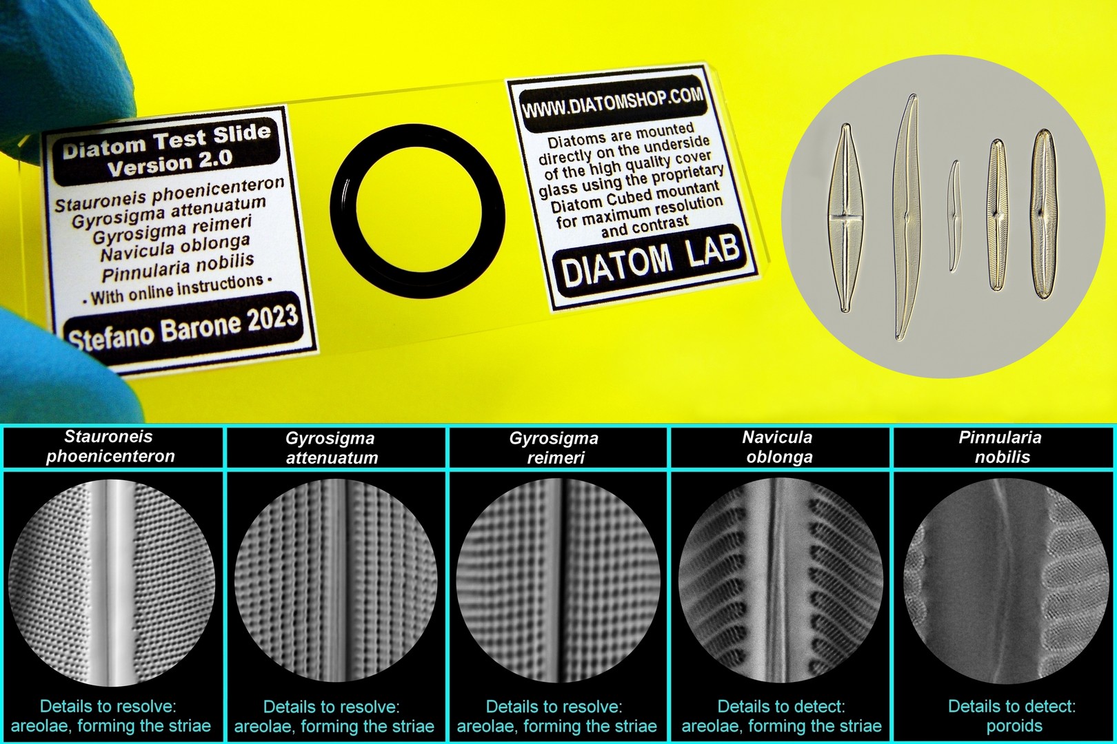
Diatom Test Slide version 2.0. Download the free Instruction manual (Updated edition) by the website! The Diatom Test Slide version 2.0 groups Test Diatoms ranging from easy to extremely difficult. Since its appearance in 2018, hundreds of laboratories, microscope companies and microscopists from all over the world use this standardized slide with profit and satisfaction! NOT ALL Pinnularia nobilis (Ehrenberg) Ehrenberg ARE THE SAME! The MOST DIFFICULT Pinnularia nobilis (Ehrenberg) Ehrenberg sample among all the others we keep was chosen for the Diatom Test Slide version 2.0 (measurements were performed using a scanning electron microscope or SEM), in fact not all Pinnularia nobilis (Ehrenberg) Ehrenberg in the world have the same poroids measurements!
249.00 €
Add
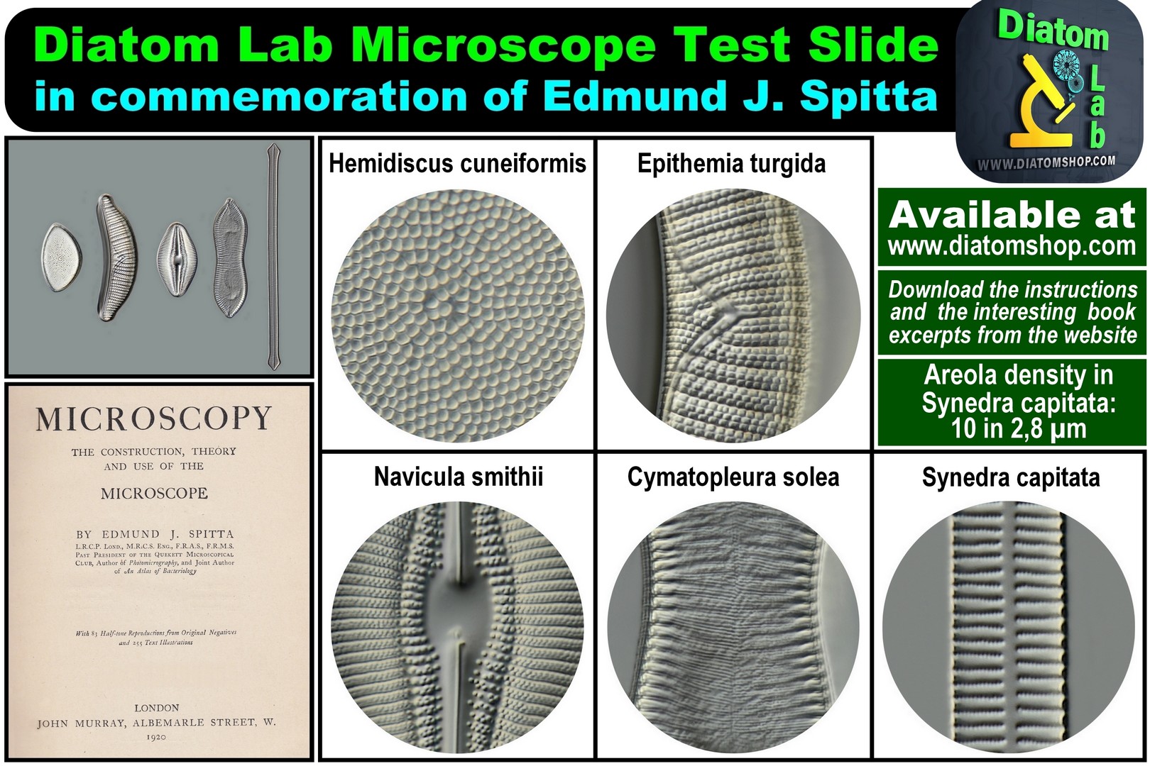
Diatom Lab Microscope Test Slide in commemoration of Edmund J. Spitta. A Complete Test for all dry and oil immersion lenses from 10x to 100x, in Bright field, Oblique illumination, Phase contrast, Darkfield, Oil immersion darkfield and DIC. Download the instructions and the interesting book excerpts from this website
249.00 €
Add
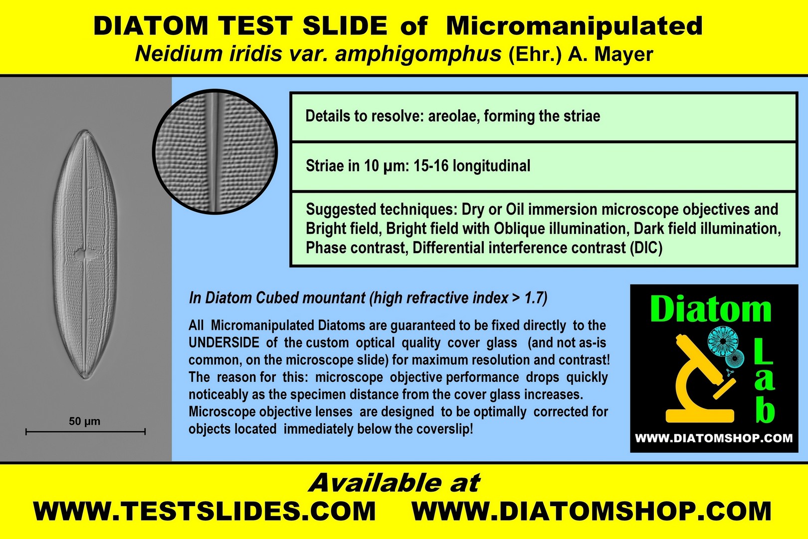
DIATOM TEST SLIDE of Micromanipulated Neidium iridis var. amphigomphus (Ehr.) A. Mayer
Details to resolve: areolae, forming the striae
Striae in 10 µm: 15-16 longitudinal
Suggested techniques: Dry or Oil immersion microscope objectives in Bright field, or Bright field with Oblique illumination, or Dark field illumination, or Phase contrast, or Differential interference contrast (DIC)
In Diatom Cubed mountant (refractive index > 1,7)
69.00 €
Add

DIATOM TEST SLIDE of micromanipulated Synedra ulna var. longissima (W.Smith) Brun 1880.
Details to resolve: areolae
Areola density: 11 in 2,8 µm
Suggested techniques: Oil immersion, double immersion. Bright field, oblique illumination,
Phase contrast, Dark field illumination, DIC
Details to resolve: areolae
Areola density: 11 in 2,8 µm
Suggested techniques: Oil immersion, double immersion. Bright field, oblique illumination,
Phase contrast, Dark field illumination, DIC
69.00 €
Add
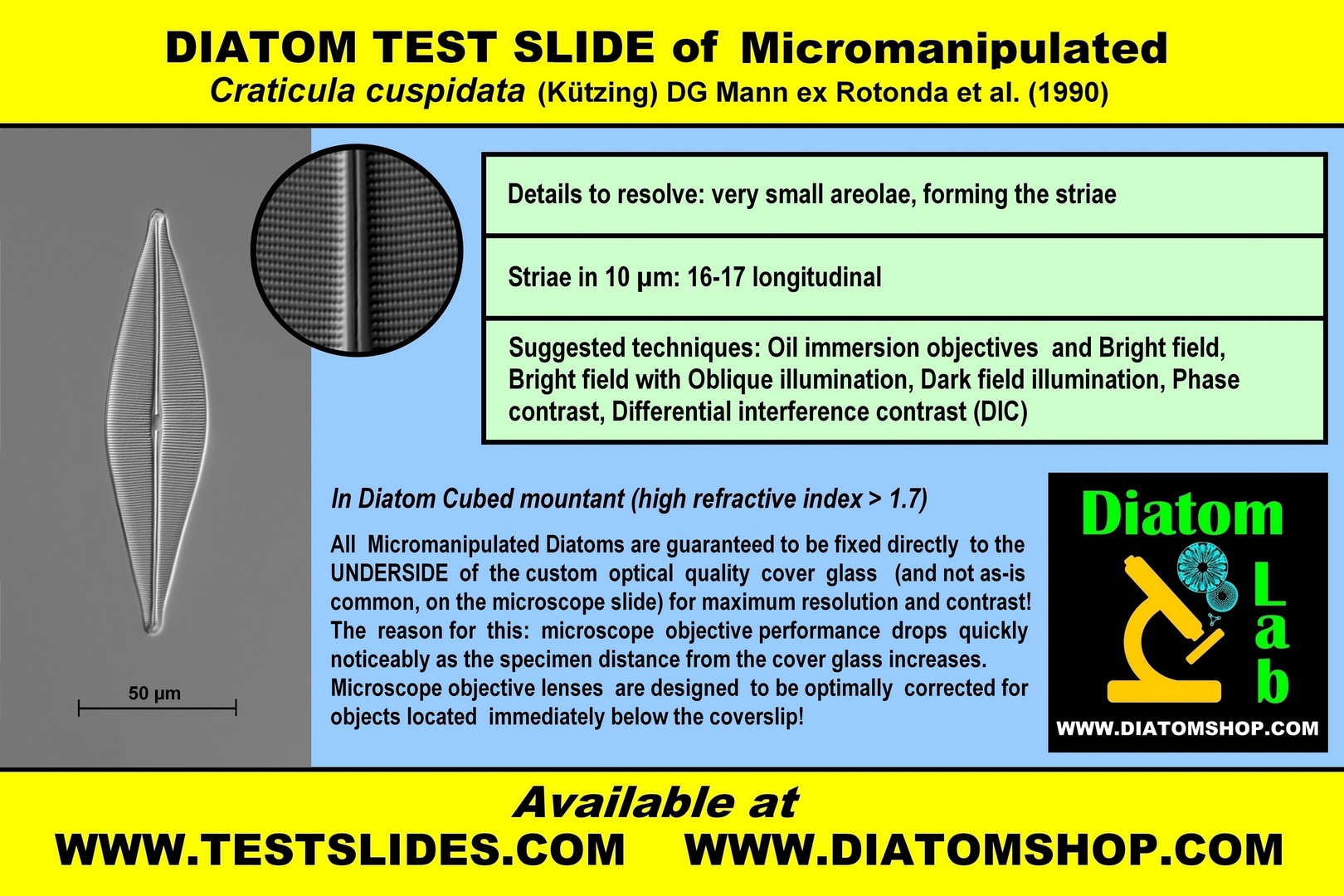
DIATOM TEST SLIDE of Micromanipulated Craticula cuspidata (Kützing) DG Mann ex Rotonda et al. (1990)
Details to resolve: very small areolae, forming the striae
Striae in 10 µm: 16-17 longitudinal
Suggested techniques: Oil immersion microscope objectives in Bright field, or Bright field with Oblique illumination, or Dark field illumination, or Phase contrast, or Differential interference contrast (DIC)
In Diatom Cubed mountant (refractive index > 1,7)
69.00 €
Add
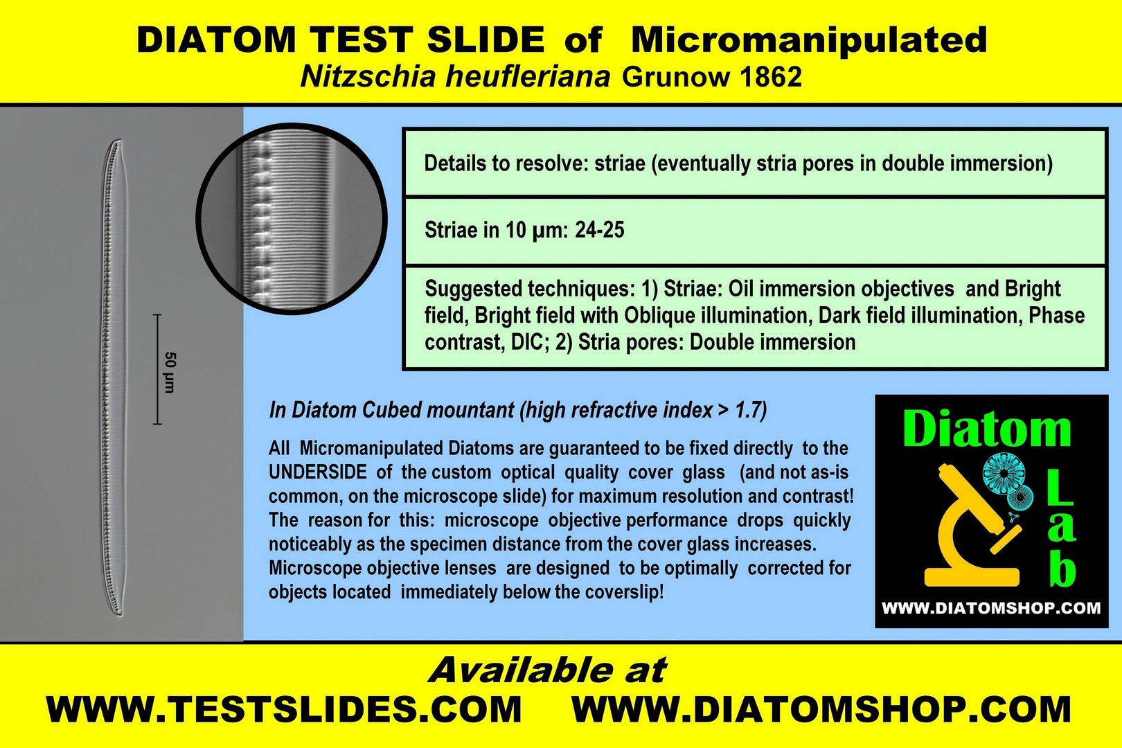
DIATOM TEST SLIDE of Micromanipulated Nitzschia heufleriana Grunow 1862
Details to resolve: striae or eventually stria pores in double immersion
Striae in 10 µm: 24-25
Suggested techniques:
1) Striae: Oil immersion microscope objectives in Bright field, or Bright field with Oblique illumination, or Dark field illumination, or Phase contrast, or Differential interference contrast (DIC);
2) Stria pores: Double immersion (= Oil immersion objective and Oil immersion condenser)
In Diatom Cubed mountant (refractive index > 1,7)
69.00 €
Add
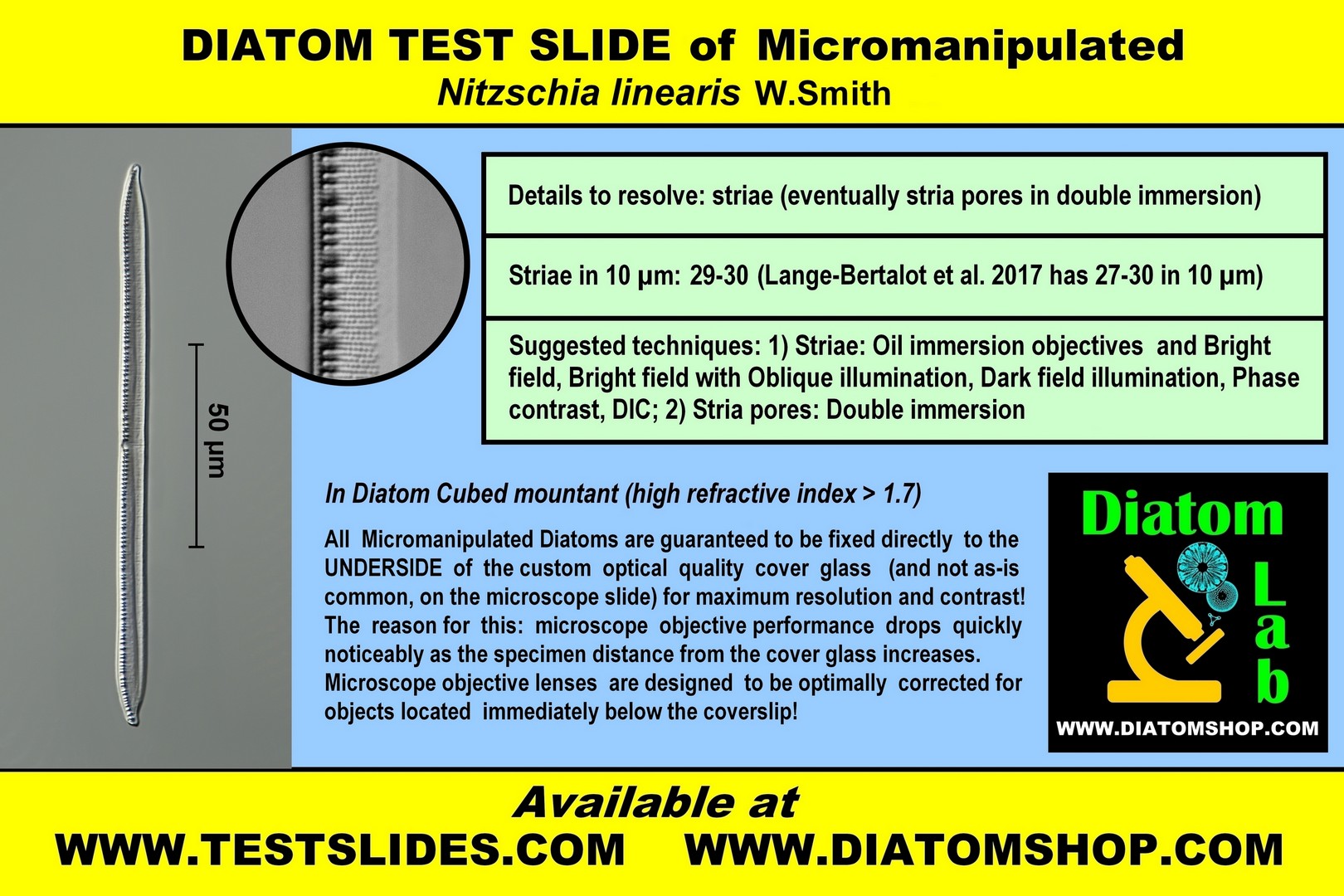
DIATOM TEST SLIDE of Micromanipulated Nitzschia linearis W.Smith
Details to resolve: striae or eventually stria pores in double immersion
Striae in 10 µm: 29-30, parallel
Suggested techniques:
1) Striae: Oil immersion microscope objectives in Bright field, or Bright field with Oblique illumination, or Dark field illumination, or Phase contrast, or Differential interference contrast (DIC);
2) Stria pores: Double immersion (= Oil immersion objective and Oil immersion condenser)
In Diatom Cubed mountant (refractive index > 1,7)
69.00 €
Add
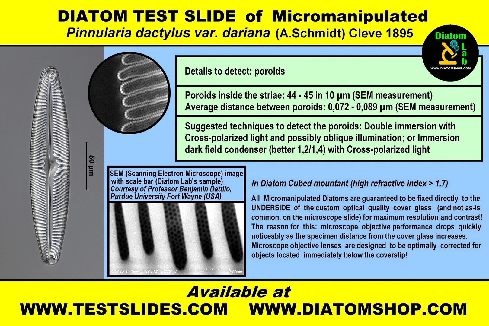
DIATOM TEST SLIDE of Micromanipulated Pinnularia dactylus var. dariana (A.Schmidt) Cleve
Details to detect: poroids
The theoretical limit of resolution of most light microscopes is ∼ 0.2 μm, but these poroids can be detected by the techniques below, thanks to Diatom Cubed high refractive index mountant!
Viewed with SEM, QUANTITATIVE FEATURES in Diatom Lab's sample are:
Poroids inside the striae: 44 - 45 in 10 µm
Average distance between poroids: 0,072 - 0,089 µm. See SEM image from the same sample!
(While In Amphipleura pellucida striae number 37-45 in 10 µm and pores are spaced 0,16 - 0,19 µm apart)
Recommended microscope objectives: Oil-immersion 63 or 100x objectives having a very good or excellent numerical aperture (1,3 or 1,4);
Suggested techniques to detect the poroids: Double immersion (= Oil immersion objective and Oil immersion condenser) with Polarized light (the polarizers should be oriented perpendicular to each other = maximum level of extinction) and possibly oblique illumination; or Immersion dark field condenser (better 1,2/1,4) with Polarized light (the polarizers should be oriented perpendicular to each other = maximum level of extinction)
In Diatom Cubed mountant (refractive index > 1,7)
79.00 €
Add
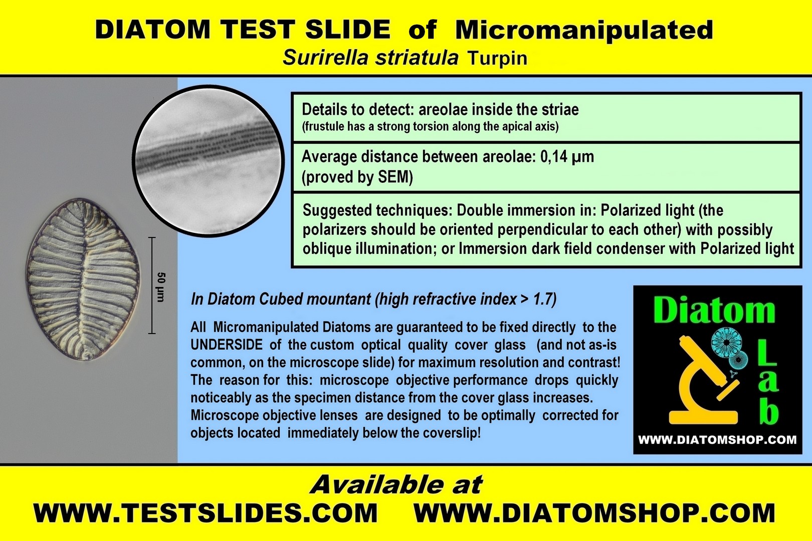
DIATOM TEST SLIDE of Micromanipulated Surirella striatula Turpin (frustule has a strong torsion along the apical axis)
Details to detect: areolae, forming the striae
Viewed with SEM, the average distance between areolae is 0,14 µm.
The theoretical limit of resolution of most light microscopes is ∼ 0.2 μm, but these areolae can be detected by the techniques below, thanks to Diatom Cubed high refractive index mountant!
Recommended microscope objectives: oil-immersion 63 or 100x objectives having a very good or excellent numerical aperture (1,3 or 1,4)
Suggested techniques: Double immersion (= Oil immersion objective and Oil immersion condenser) in: Polarized light (the polarizers should be oriented perpendicular to each other = maximum level of extinction) with possibly oblique illumination; or Immersion dark field condenser (better 1,2/1,4) with Polarized light (the polarizers should be oriented perpendicular to each other = maximum level of extinction)
In Diatom Cubed mountant (refractive index > 1,7)
69.00 €
Add
OPTICAL ABERRATIONS DETECTOR. It's a very useful Diatom Lab microscope slide that detects optical aberrations - even the most imperceptible ones - in light microscopy, such as: a) Curvature of field (the image appears sharp and crisp in the center or on the edges of the viewfield but not both); b) Distortion (changes in the shape of the image); c) Barrel distortion and Pincushion distortion; d) Spherical aberration (the specimen image appears hazy or blurred and slightly out of focus); e) Chromatic aberration (presence of color fringes at the edges of the borders); f) Astigmatism (the ideal circular point image blurs into a diffuse circle, elliptical patch, or line). Thanks to this unique microscope slide, you can easily test and compare your objectives, eyepieces, condensers, phototubes, microscope cameras, and magnification changers! This is an amazing microscope slide in the center of which there are, incredibly, 90.000 microscopic perfect squares having a side of 10 µm (ten micrometres or microns) and 10 µm apart! These microscopic perfect squares also form nine larger squares useful in case of low magnification (see the page "Optical Aberrations Detector, Centering Device, Micrometers" for better image quality). This slide is excellent for testing macro lenses as well! Click on this link to download the pdf file. All Rights Reserved.
Optical Aberrations Detector for transmitted light (with cover glass and mountant).
249.00 €
Add
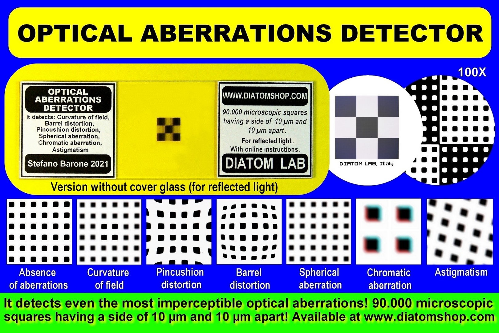
Optical Aberrations Detector for reflected light (without cover glass).
249.00 €
Add
CENTERING DEVICE & MICROMETER GLASS SLIDE. It's an all-in-one microscope slide that incorporates a centering device to center the microscope and the camera and a stage micrometer for microscope calibration. The centering device consists of 25 perfect concentric circles, 50 µm (fifty micrometres or microns) thick and 50 µm apart! It is very useful to center the microscope parts (condenser top lens, field iris diaphragm, prisms of the binocular/trinocular head, etc.) and the microscope camera. (It is advisable to make changes to the microscope only if you are an expert.) The stage micrometer for microscope calibration is a 1 mm Scale with 0,01 mm (10 µm) divisions; the slide can be used in combination with a micrometer eyepiece or a measurement software (for cameras) in order to measure preparations. (See the page "Optical Aberrations Detector, Centering Device, Micrometers" for better image quality.) All Rights Reserved.
For transmitted light (with cover glass and mountant). Diatom Lab CENTERING DEVICE & MICROMETER GLASS SLIDE is an all-in-one microscope slide that incorporates a centering device to center the microscope and the camera and a stage micrometer for microscope calibration. The centering device consists of 25 perfect concentric circles, 50 µm (fifty micrometres or microns) thick and 50 µm apart! It is very useful to center the microscope parts (condenser top lens, field iris diaphragm, prisms of the binocular/trinocular head, etc.) and the microscope camera. The stage micrometer for microscope calibration is a 1 mm Scale with 0,01 mm (10 µm) divisions. The slide can be used in combination with a micrometer eyepiece or a measurement software (for cameras) in order to measure preparations.
99.00 €
Add
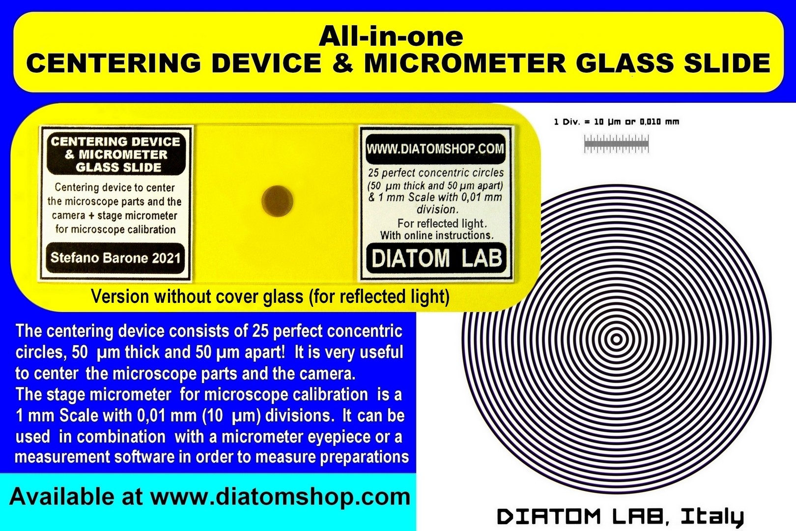
For reflected light (without cover glass). Diatom Lab CENTERING DEVICE & MICROMETER GLASS SLIDE is an all-in-one microscope slide that incorporates a centering device to center the microscope and the camera and a stage micrometer for microscope calibration. The centering device consists of 25 perfect concentric circles, 50 µm (fifty micrometres or microns) thick and 50 µm apart! It is very useful to center the microscope parts (condenser top lens, field iris diaphragm, prisms of the binocular/trinocular head, etc.) and the microscope camera. The stage micrometer for microscope calibration is a 1 mm Scale with 0,01 mm (10 µm) divisions. The slide can be used in combination with a micrometer eyepiece or a measurement software (for cameras) in order to measure preparations.
99.00 €
Add
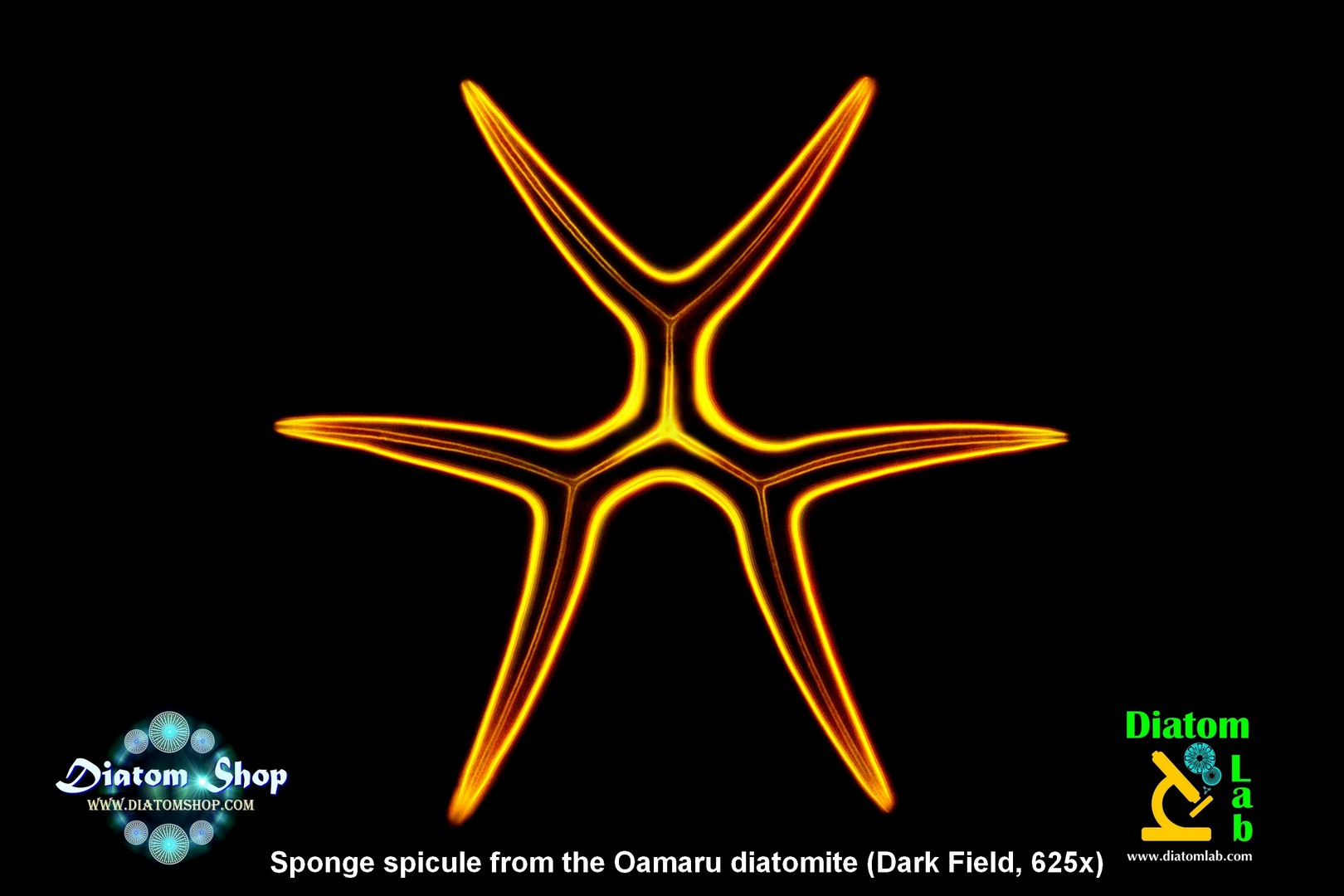
STREW DIATOM SLIDES: enjoy the DIATOM SAFARI Each microscope slide contains dozens, if not hundreds, of strewn cleaned Diatoms that haven't been selected and micromanipulated. The Diatoms in the images below are just an example of the species you might find in each slide, in fact the species list of each sample is copious (the more slides you buy, the more chance you have of finding different species). Strew slides reveal the complex nature of a sample at a glance: unlike slides containing selected and micromanipulated Diatoms (see further down), some strew slides may also contain few radiolarians, diatom fragments and spicules. All strewn Diatoms are guaranteed to be fixed directly to the UNDERSIDE of the custom optical quality cover glass (and not, as is common, on the microscope slide) for maximum resolution and contrast! The reason for this: microscope objective performance drops quickly noticeably as the specimen distance from the cover glass increases. Microscope objective lenses are designed to be optimally corrected for objects located immediately below the coverslip!

NEW! VERY RARE! Strew microscope slide of recent freshwater Surirella biseriata var. ianus v. nov. + Surirella biseriata f. constricta + Surirella biseriata, all from a natural sample collected in Bavarian Forest (Germany)! Read the interesting informations on the side. The sample is made up of 99% Surirella diatoms! Each strew slide contains dozens of largely intact specimens, excellent for exciting observations and superb microscope images! A unique microscope slide.
129.00 €
Add
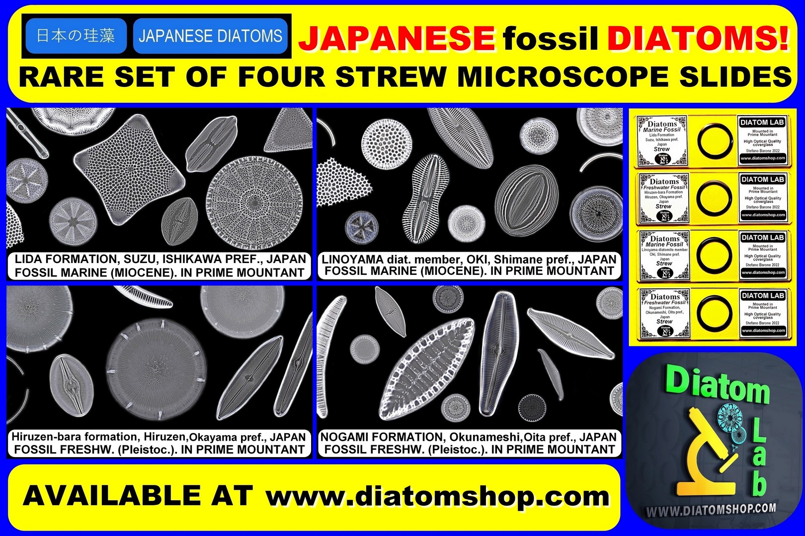
NEW! RARE set of four strew microscope slides: RARE fossil Japanese Diatoms from four locations! All samples contains many species (images in dark field are just examples of what you can find). RARE SPECIES INCLUDE: Arachnoidiscus sendaicus N.E.Brown, 1933 (Suzu sample), Rutilaria longicornis Tempère & Brun in Brun & Tempère, 1889 (Suzu sample), Denticulopsis praehyalina Tanimura 1989 (Oki sample), Diploneis yatukaensis Horikawa & Okuno in Okuno, 1944 (Hiruzen sample), Didymosphenia fossilis Horikawa & Okuno, 1944 (Okunameshi sample), Pinnularia hartleyana var. notata Kobayashi 1968 (Okunameshi sample)... AND MANY MORE!
99.00 €
Add
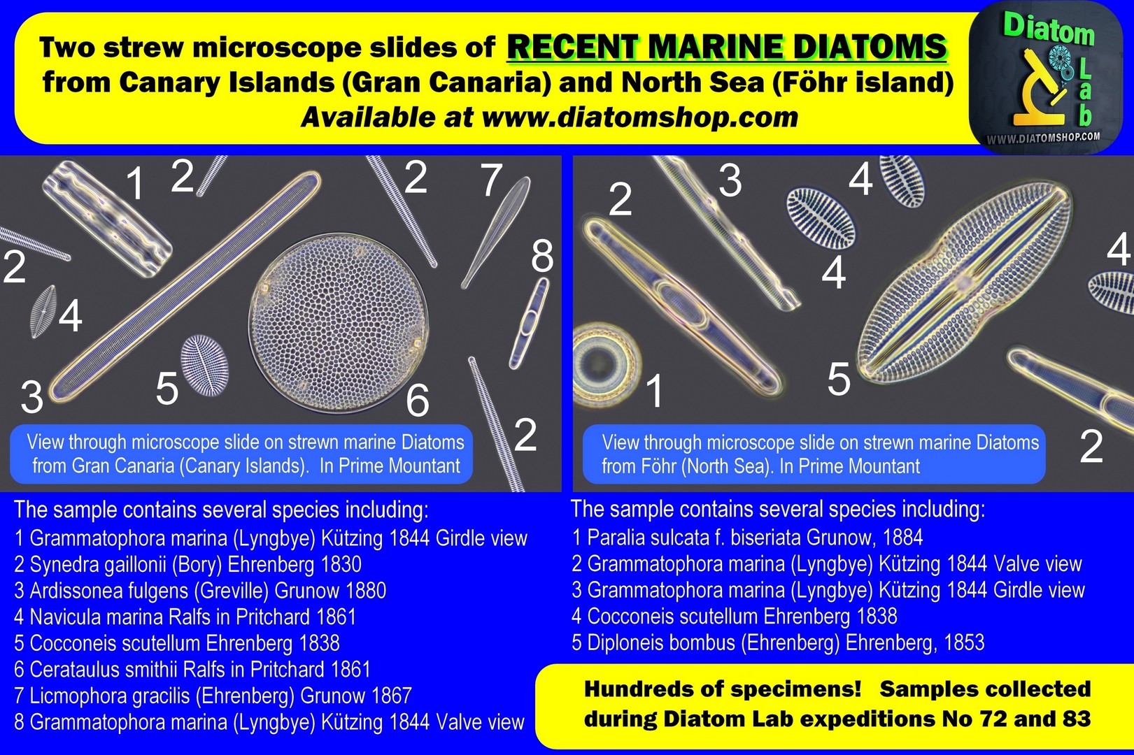
Two strew microscope slides of recent marine Diatoms from Canary Islands and North Sea. The more you buy, the more chance you have of finding different species! Strewn Diatoms are mounted in Prime Mountant (excellent high refractive index mountant with refractive index > 1,7)
79.00 €
Add
The more you buy, the more chance you have of finding different species!
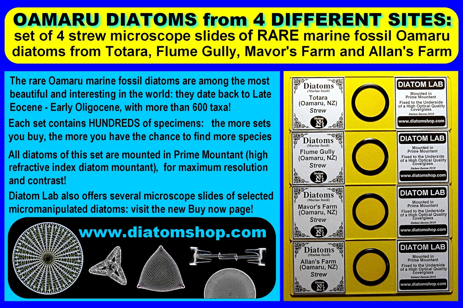
Set of 4 strew microscope slides of RARE marine fossil OAMARU diatoms from 4 different sites: Totara, Flume Gully, Mavor's Farm, Allan's Farm. Hundred of specimens! Strewn Diatoms are mounted in Prime Mountant (excellent high refractive index mountant with refractive index > 1,7), with hundreds of specimens
Set of 4 strew microscope slides of RARE marine fossil OAMARU diatoms from 4 different sites: Totara, Flume Gully, Mavor's Farm, Allan's Farm. Hundred of specimens! Strewn Diatoms are mounted in Prime Mountant (excellent high refractive index mountant with refractive index > 1,7), with hundreds of specimens
99.00 €
Add
The more you buy, the more chance you have of finding different species!
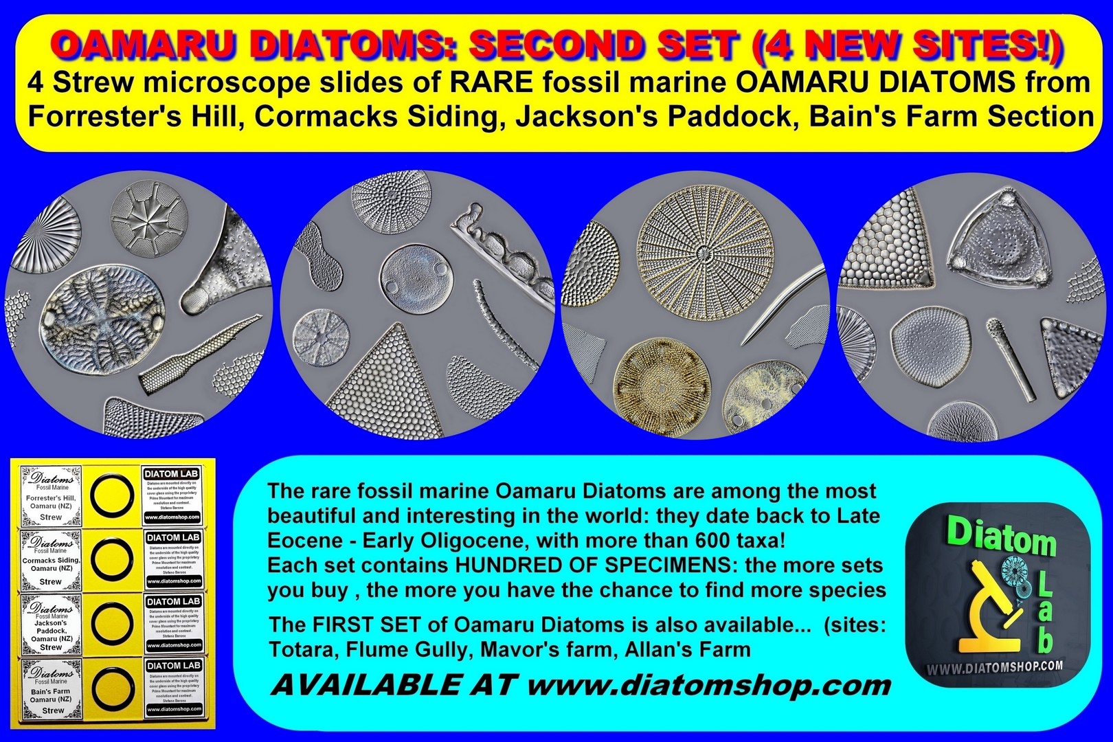
Set of 4 strew microscope slides of RARE marine fossil OAMARU diatoms from 4 different sites: Forrester's Hill, Cormacks Siding, Jackson's Paddock, Bain's Farm Section. Hundred of specimens! Strewn Diatoms are mounted in Prime Mountant (excellent high refractive index mountant with refractive index > 1,7), with hundreds of specimens
Set of 4 strew microscope slides of RARE marine fossil OAMARU diatoms from 4 different sites: Forrester's Hill, Cormacks Siding, Jackson's Paddock, Bain's Farm Section. Hundred of specimens! Strewn Diatoms are mounted in Prime Mountant (excellent high refractive index mountant with refractive index > 1,7), with hundreds of specimens
99.00 €
Add
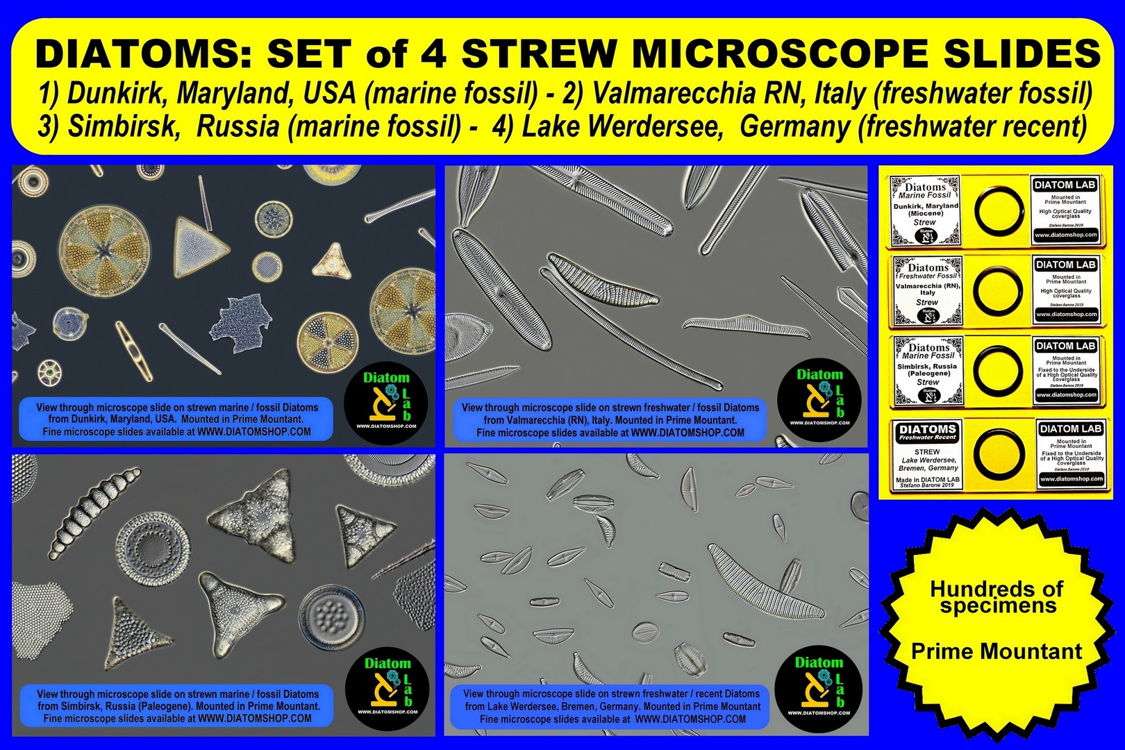
Set of 4 strew microscope slides of Diatoms from Dunkirk, Maryland, USA; Valmarecchia, RN, Italy; Simbirsk, Russia; Lake Werdersee, Germany. The more you buy, the more chance you have of finding different species!Strewn Diatoms are mounted in Prime Mountant (excellent high refractive index mountant with refractive index > 1,7)
99.00 €
Add
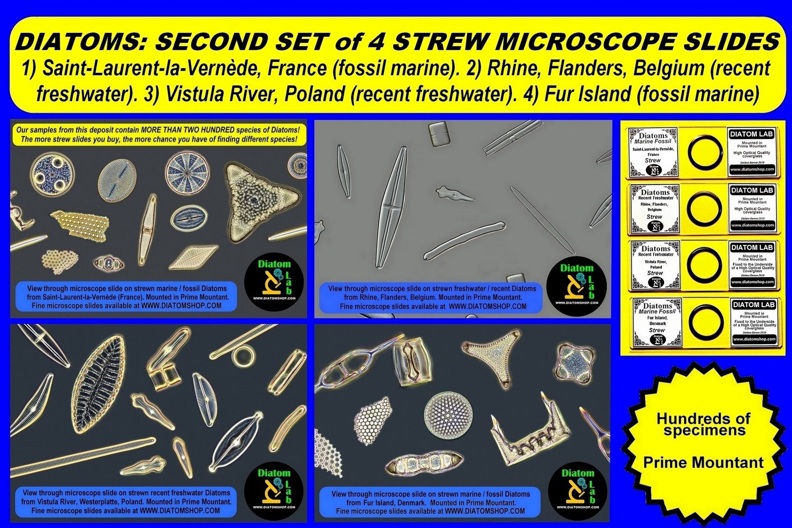
Set of 4 strew microscope slides of Diatoms from Saint-Laurent-la-Vernède, France; Rhine, Flanders, Belgium; Vistula River, Poland; Fur Island, Denmark. The more you buy, the more chance you have of finding different species!Strewn Diatoms are mounted in Prime Mountant (excellent high refractive index mountant with refractive index > 1,7)
99.00 €
Add
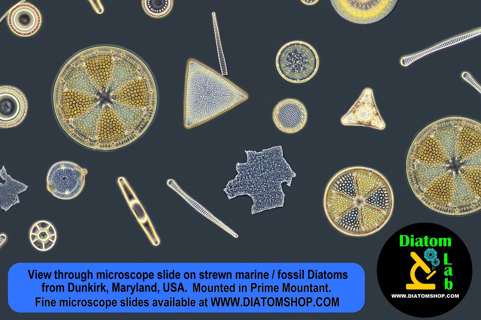
Strew microscope slide of Diatoms from Dunkirk, Maryland, USA, sample B (marine fossil, Miocene). The more you buy, the more chance you have of finding different species!Strewn Diatoms are mounted in Prime Mountant (excellent high refractive index mountant with refractive index > 1,7)
29.00 €
Add
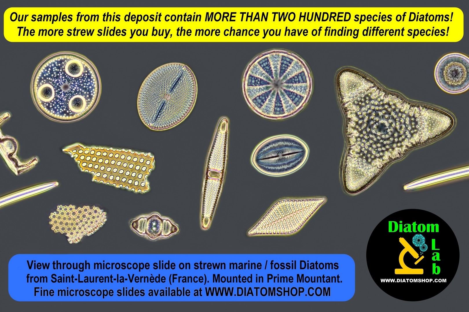
Strew microscope slide of Diatoms from Saint Laurent La Vernede, France (marine fossil, early Miocene). The more you buy, the more chance you have of finding different species!Strewn Diatoms are mounted in Prime Mountant (excellent high refractive index mountant with refractive index > 1,7)
29.00 €
Add
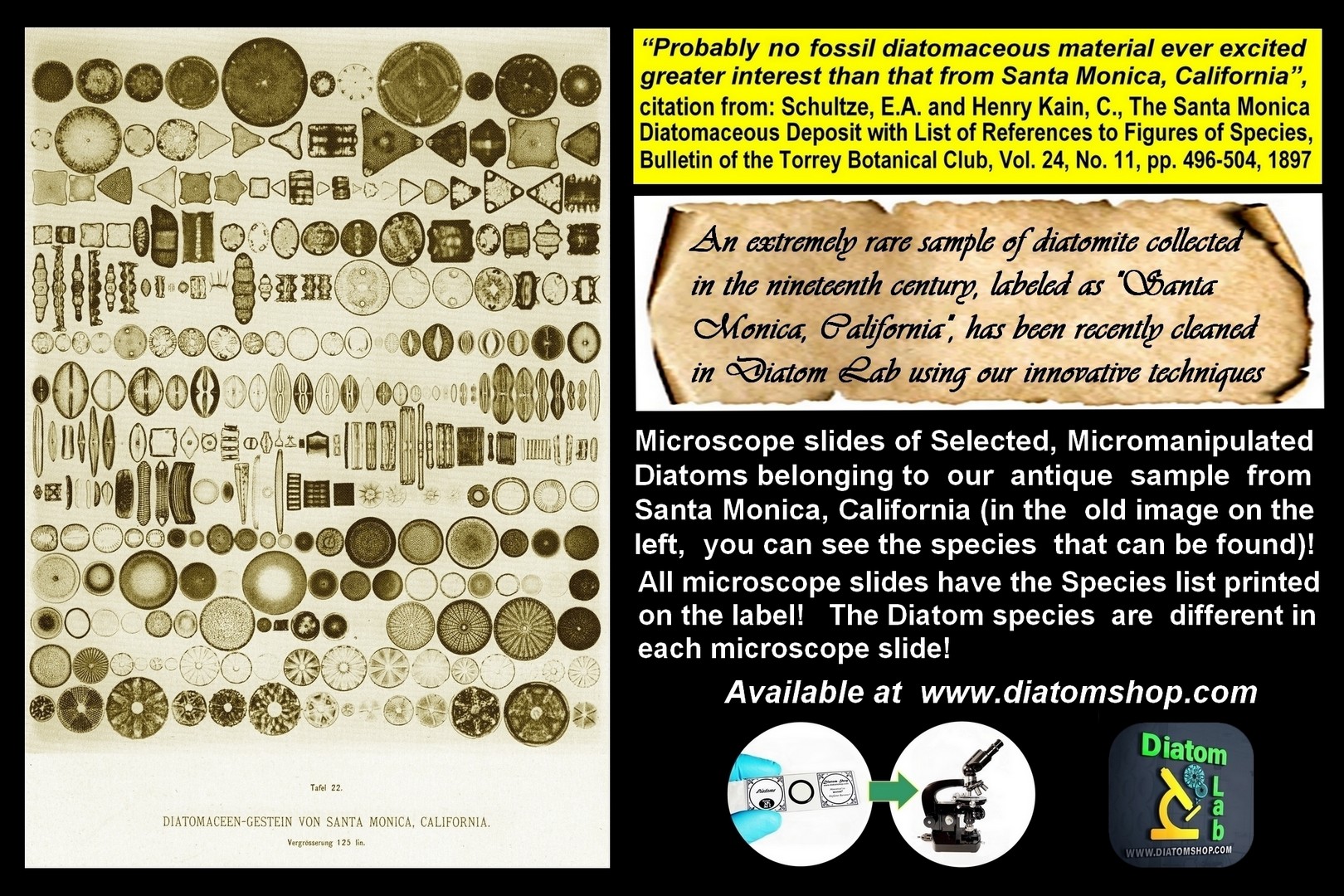
“Probably non fossil diatomaceous material ever excited greater interest than that from Santa Monica, California”, citation from: Schultze, E.A. and Henry Kain, C., The Santa Monica Diatomaceous Deposit with List of References to Figures of Species, Bulletin of the Torrey Botanical Club, Vol. 24, No. 11, pp. 496-504, 1897. The more you buy, the more chance you have of finding different species!Strewn Diatoms are mounted in Prime Mountant (excellent high refractive index mountant with refractive index > 1,7)
29.00 €
Add
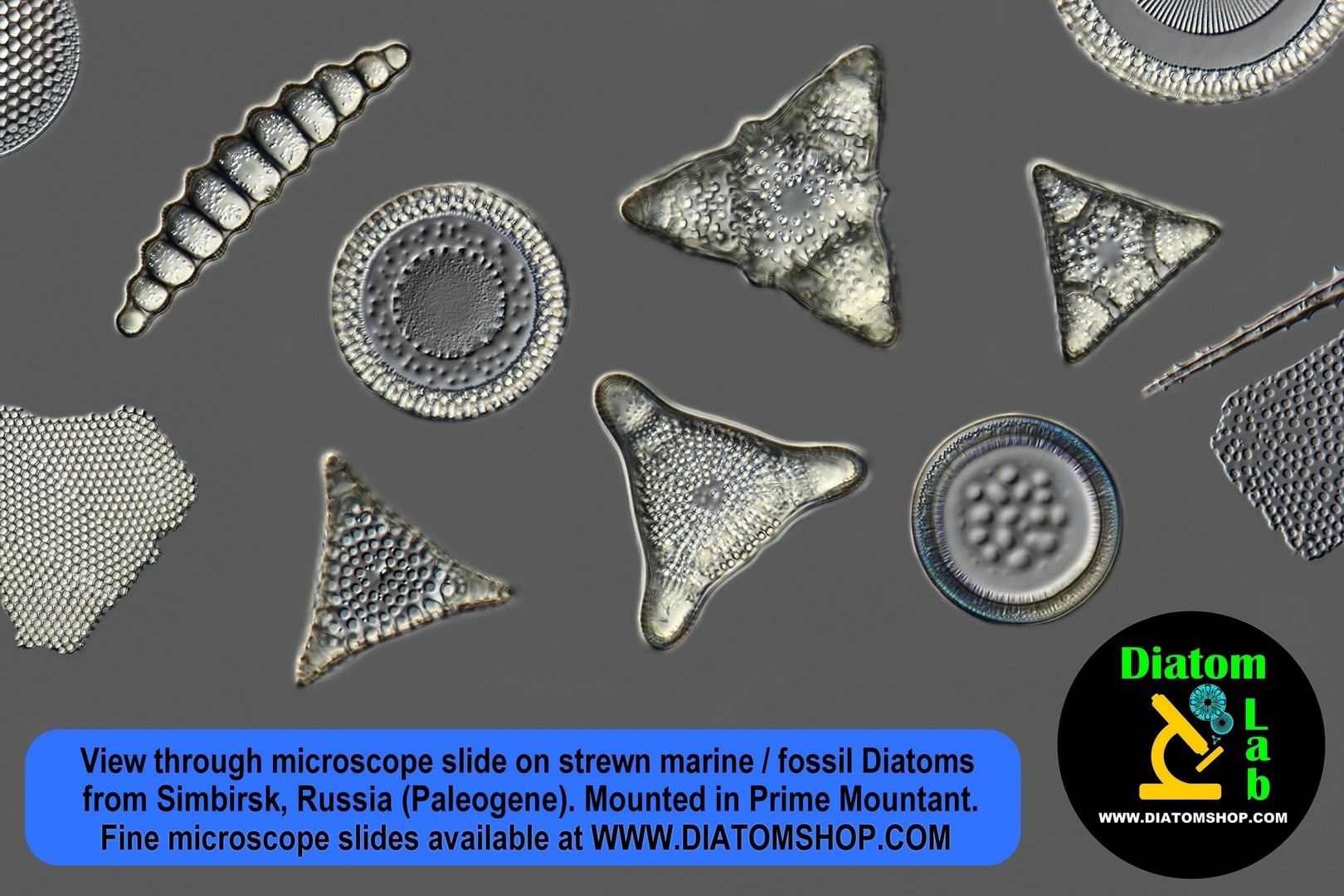
Strew microscope slide of Diatoms from Simbirsk, Russia (marine fossil, Paleogene). RARE SAMPLE! The more you buy, the more chance you have of finding different species!Strewn Diatoms are mounted in Prime Mountant (excellent high refractive index mountant with refractive index > 1,7)
39.00 €
Add
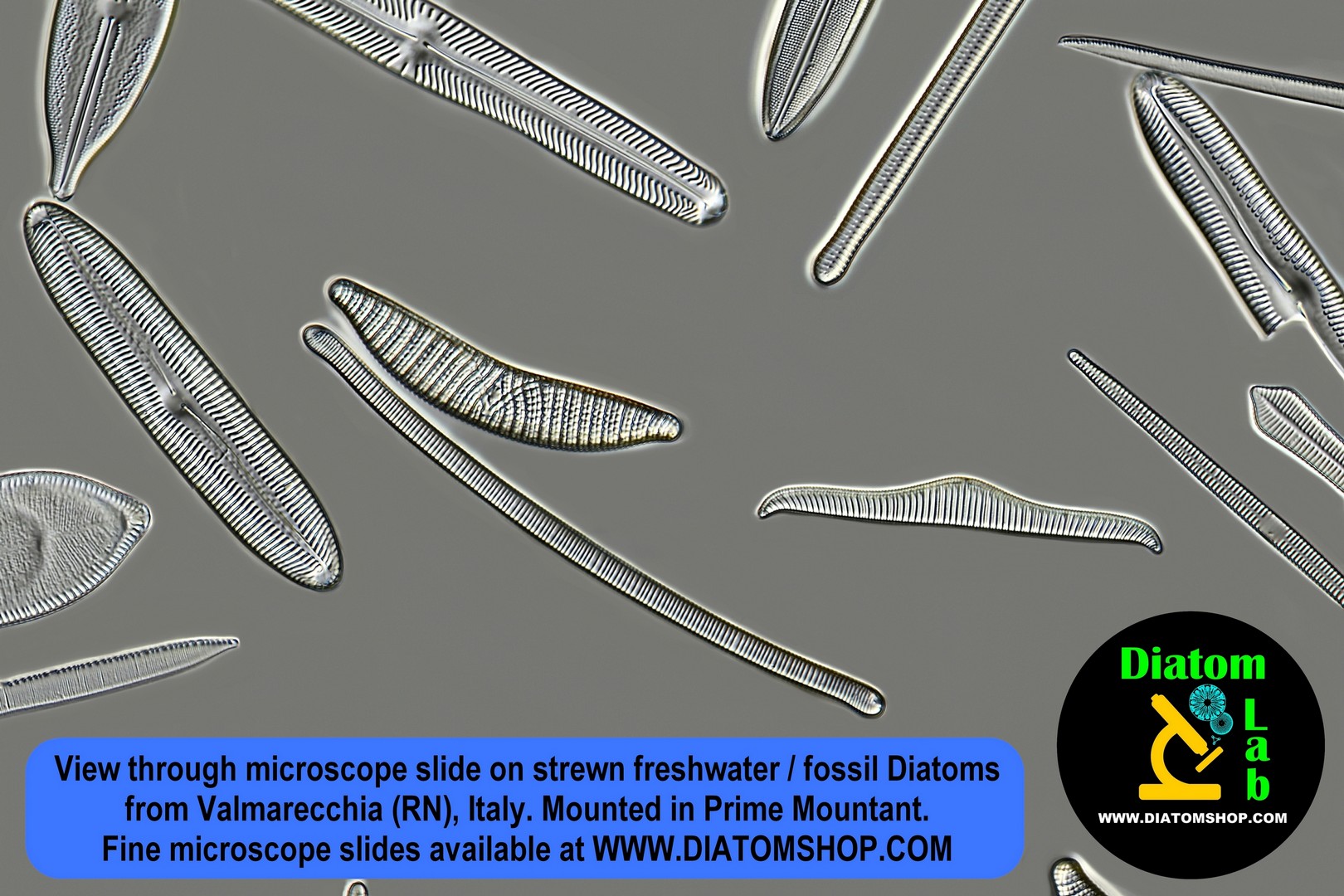
Strew microscope slide of Diatoms from Valmarecchia(RN), Italy (freshwater fossil). RARE SMPLE! The more you buy, the more chance you have of finding different species!Strewn Diatoms are mounted in Prime Mountant (excellent high refractive index mountant with refractive index > 1,7)
29.00 €
Add
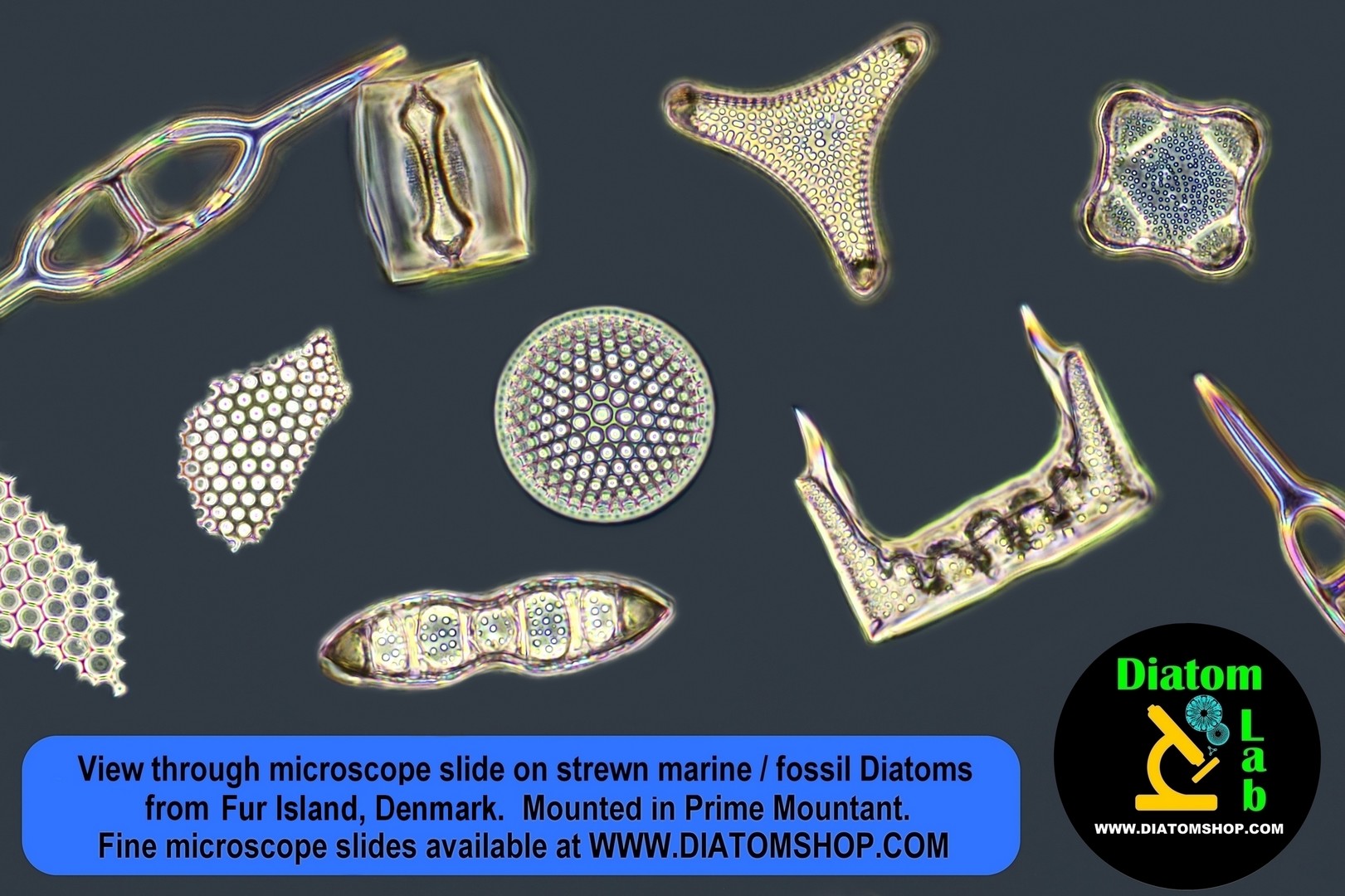
Strew microscope slide of Diatoms from Fur Island, Denmark (marine fossil, Paleogene). The more you buy, the more chance you have of finding different species!Strewn Diatoms are mounted in Prime Mountant (excellent high refractive index mountant with refractive index > 1,7)
29.00 €
Add
Strew microscope slide of Diatoms from Oamaru, Allan's Farm (marine fossil, late Eocene to early Oligocene). The more you buy, the more chance you have of finding different species!Strewn Diatoms are mounted in Prime Mountant (excellent high refractive index mountant with refractive index > 1,7)
39.00 €
Add
MICROSCOPE SLIDES OF SELECTED, MICROMANIPULATED DIATOMS. All micromanipulated Diatoms are guaranteed to be fixed directly to the UNDERSIDE of the custom, optical quality cover glass (and not, as is common, on the microscope slide) for maximum resolution and contrast! The reason for this is that: microscope objective performance drops quickly and noticeably as the specimen distance from the cover glass increases. Microscope objective lenses are designed to be optimally corrected for objects located immediately below the coverslip!

VERY RARE! Microscope slide of Selected, Micromanipulated Surirella biseriata v. ianus v. nov. (more informations on the side) + Surirella biseriata f. constricta + S. biseriata, from a unique, amazing sample collected in Bavarian Forest (Germany). A scientific unicum.
249.00 €
Add
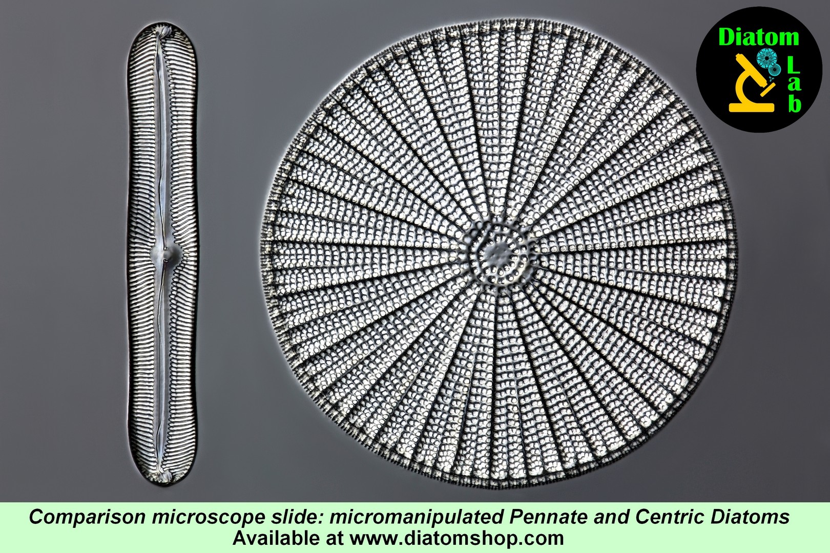
Microscope slide of one micromanipulated Pennate Diatom (bilaterally symmetric) and one micromanipulated Centric Diatom (radially symmetric), mounted in the excellent Diatom Cubed mountant (refractive index > 1,7). See example in DIC on the side.
59.00 €
Add

Microscope slide of 10 selected, micromanipulated fossil marine Diatoms from Sisquoc Formation, Lompoc, Ca, USA (Miocene), with Species List printed on the label. In Diatom Cubed mountant. Among the most beautiful and interesting species we mention: Diploneis gemmata (Greville) Cleve, 1894; Stictodiscus kittonianus Greville 1861; Stictodiscus californicus f. ecostata Grunow in Schmidt et al., 1882; Navicula praetexta f. praetexta Ehrenberg, 1840; Auliscus punctatus Bailey. The species photographed here are an example of those you might find, in fact our samples contain many species (every microscope slide is unique)
149.00 €
Add
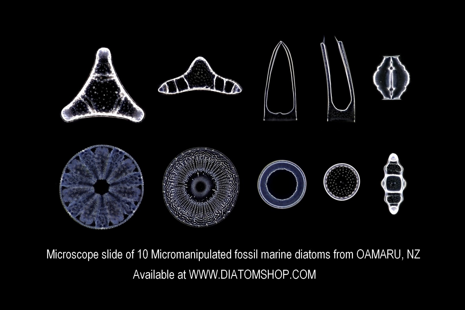
Microscope slide of 10 Selected mixed fossil marine diatoms from OAMARU, NZ (Upper Eocene Epoch: from 37.2 +/- 0.1 To 33.9+/- 0.1 million years ago), with Species list printed on the label. Micromanipulated in parallel lines (without duplicates) and mounted in the excellent Diatom Cubed mountant (refractive index > 1,7). See example in dark field on the side: this sample contains many more species!
169.00 €
Add

Microscope slide of 10 Very Rare, Micromanipulated Japanese diatoms from Suzu (fossil marine specimens, Lida formation, Miocene), with Species List printed on the label. Micromanipulated in parallel lines (without duplicates) and mounted in the excellent Diatom Cubed mountant (refractive index > 1,7). See example in dark field illumination on the side: this sample contains many more species!
169.00 €
Add
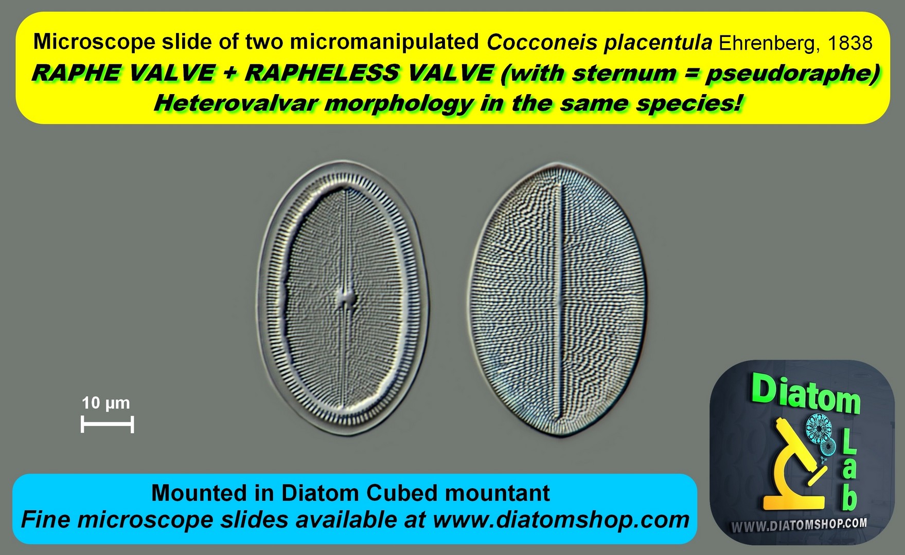
Teaching microscope slide of 2 micromanipulated Cocconeis placentula Ehrenberg, 1838 with RAPHE valve and RAPHELESS valve [exhibiting a sternum = pseudoraphe]. Cocconeis placentula has a heterovalvar morphology! Mounted in the excellent Diatom Cubed mountant (refractive index > 1,7)
99.00 €
Add
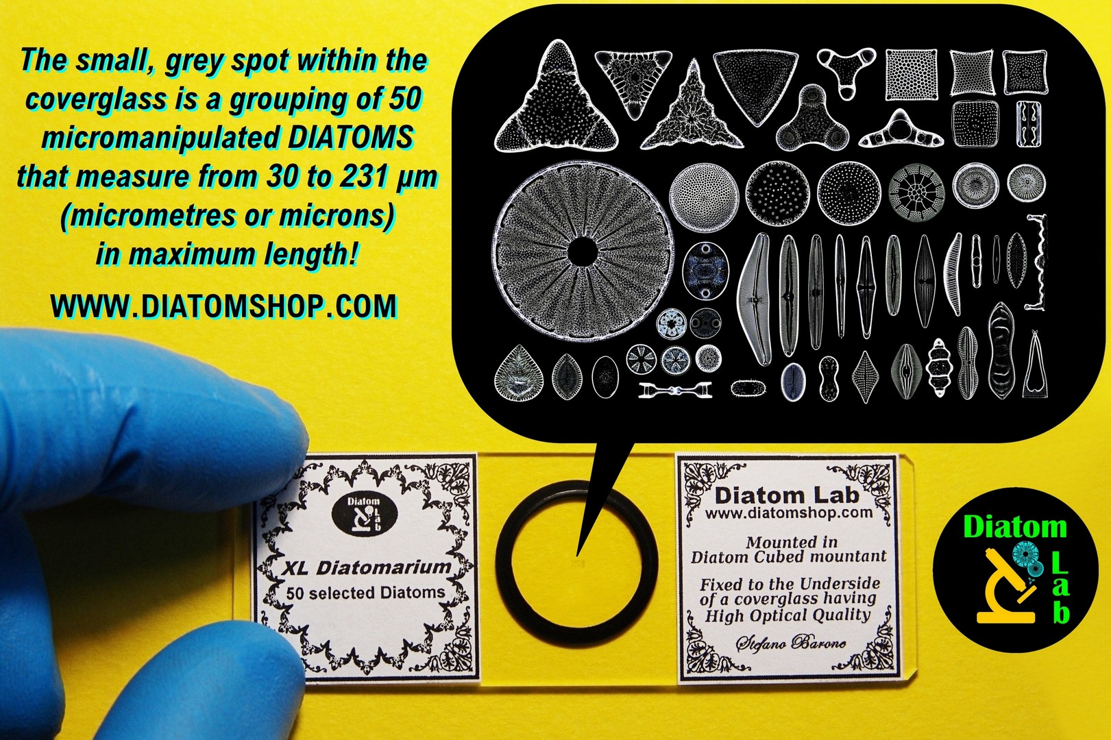
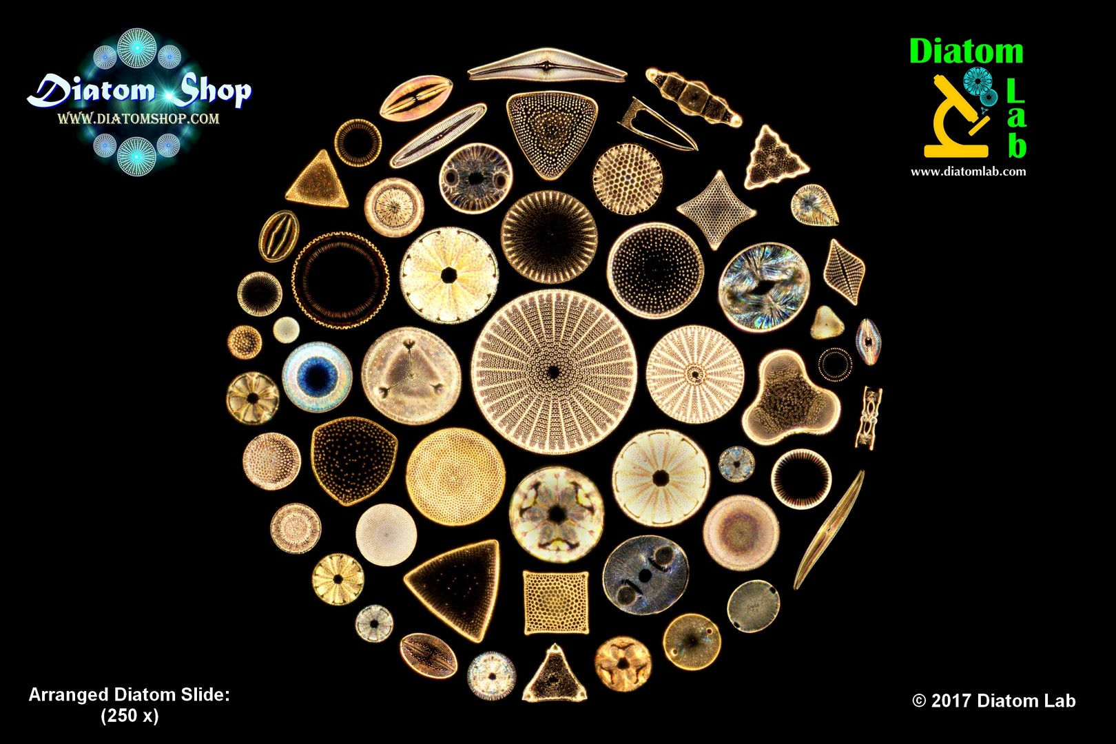
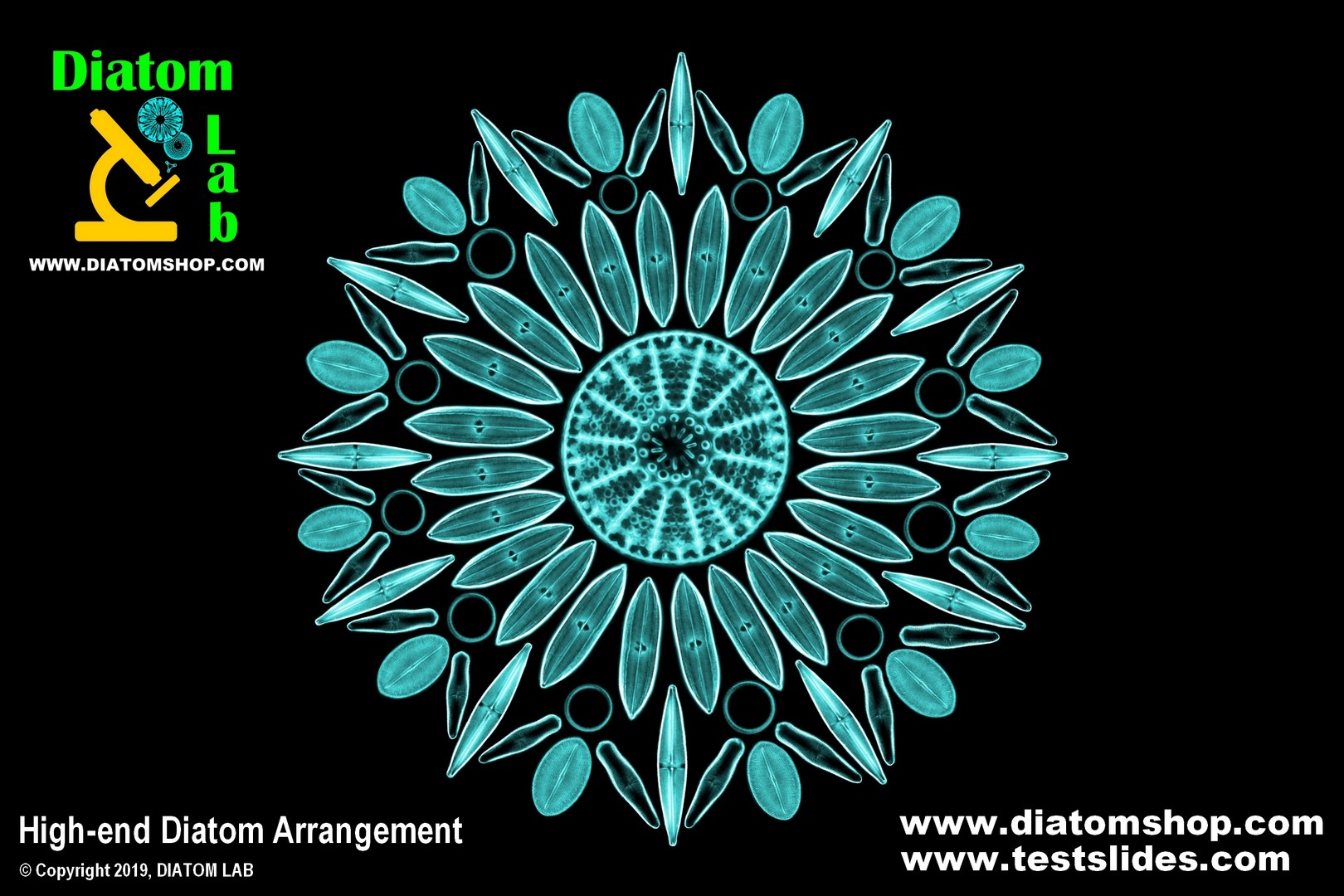
Microscope slide of 50 Selected mixed (recent - fossil - marine - freshwater) diatoms, micromanipulated in parallel lines (without duplicates), mounted in the excellent Diatom Cubed mountant (refractive index > 1,7). EACH DIATOMARIUM is a UNIQUE PIECE of MICROSCOPIC ART, as the Diatom species and the arrangement vary: in fact DIATOM LAB keeps THOUSANDS OF DIFFERENT marine / freshwater / recent / fossil / subfossil DIATOM SPECIES within the laboratory! Look at the example in dark field illumination on the side
399.00 €
Add
Microscope slide of 25 Selected mixed (recent - fossil - marine - freshwater) diatoms, micromanipulated in parallel lines (without duplicates), mounted in the excellent Diatom Cubed mountant (refractive index > 1,7). EACH DIATOMARIUM is a UNIQUE PIECE of MICROSCOPIC ART, as the Diatom species and the arrangement vary: in fact DIATOM LAB keeps THOUSANDS OF DIFFERENT marine / freshwater / recent / fossil / subfossil DIATOM SPECIES within the laboratory! Look at the example of XL Diatomarium
249.00 €
Add
Microscope slide of 16 Selected mixed (recent - fossil - marine - freshwater) diatoms, micromanipulated in parallel lines (without duplicates), mounted in the excellent Diatom Cubed mountant (refractive index > 1,7). EACH DIATOMARIUM is a UNIQUE PIECE of MICROSCOPIC ART, as the Diatom species and the arrangement vary: in fact DIATOM LAB keeps THOUSANDS OF DIFFERENT marine / freshwater / recent / fossil / subfossil DIATOM SPECIES within the laboratory! Look at the example of XL Diatomarium
169.00 €
Add
Microscope slide of 10 selected mixed (recent - fossil - marine - freshwater) diatoms, micromanipulated in parallel lines (without duplicates), mounted in the excellent Diatom Cubed mountant (refractive index > 1,7). EACH DIATOMARIUM is a UNIQUE PIECE of MICROSCOPIC ART, as the Diatom species and the arrangement vary: in fact DIATOM LAB keeps THOUSANDS OF DIFFERENT marine / freshwater / recent / fossil / subfossil DIATOM SPECIES within the laboratory! Look at the example of XL Diatomarium
149.00 €
Add
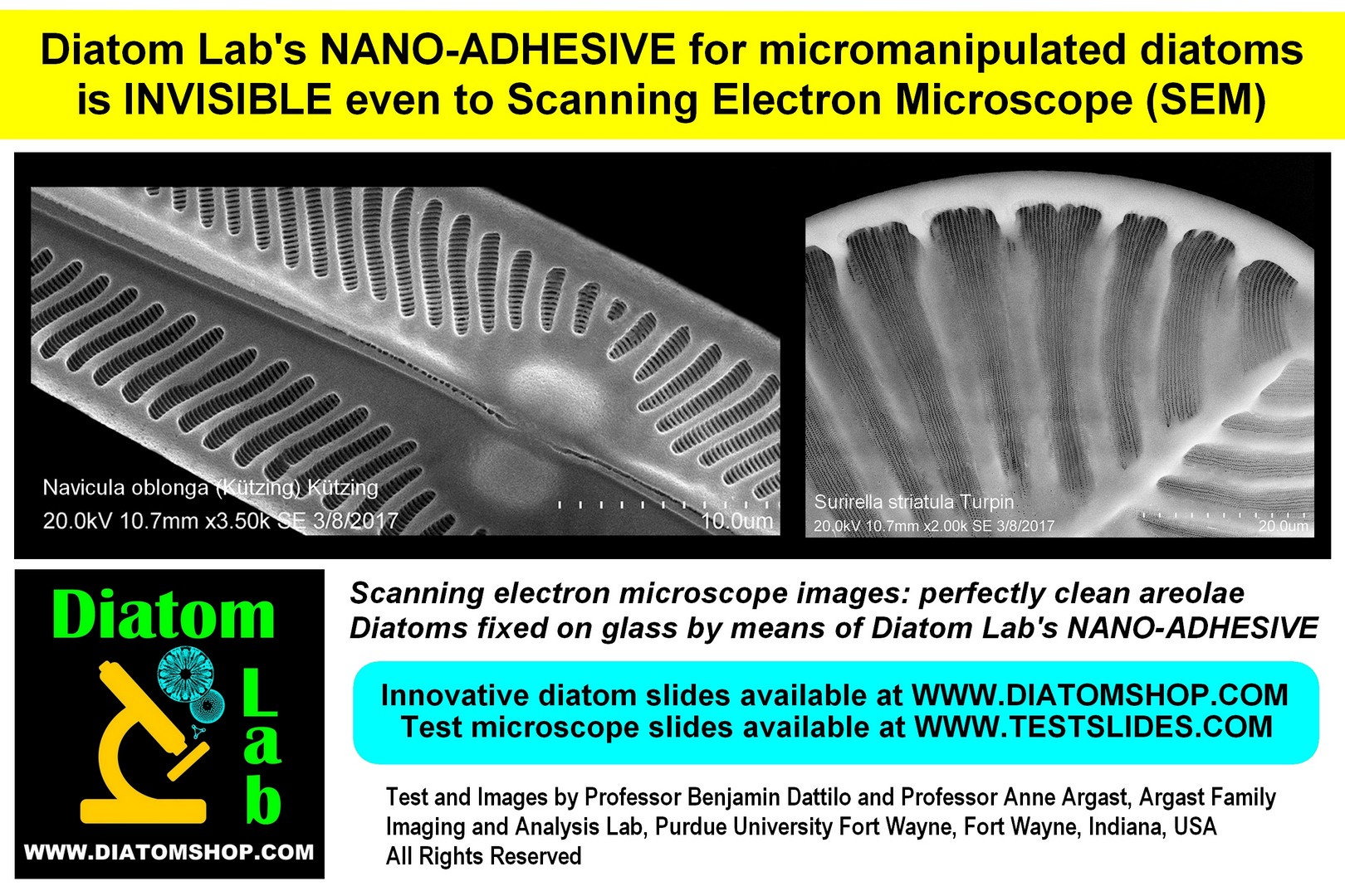
10 selected, micromanipulated Diatoms for Scanning Electron Microscope (SEM) and Atom Force Microscope (AFM), with Species list, micromanipulated and fixed on microscope slide without cover glass and mountant of course (for other quantities or specific species please ask). Diatoms are fixed by means of the proprietary Diatom Lab's NANO-ADHESIVE, which is invisible to scanning electron microscopes and is therefore used to obtain a clean background for our SEM specimen preparations!
299.00 €
Add
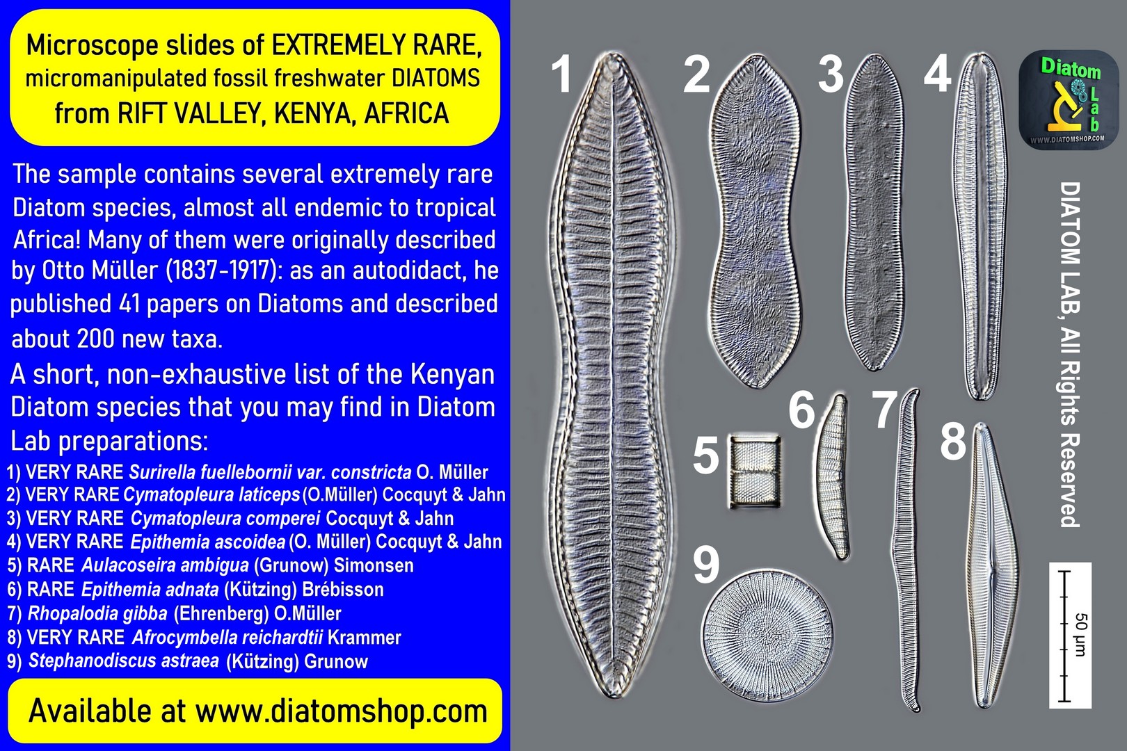
Microscope slide of 8 Very Rare, Selected, Micromanipulated fossil freshwater Diatoms from Rift Valley, Kenya, Africa, with Species list printed on the label. Micromanipulated in parallel lines (without duplicates) and mounted in the excellent Diatom Cubed mountant (refractive index > 1,7). See example of the species in DIC on the side: this Very Rare sample contains many more species!
199.00 €
Add
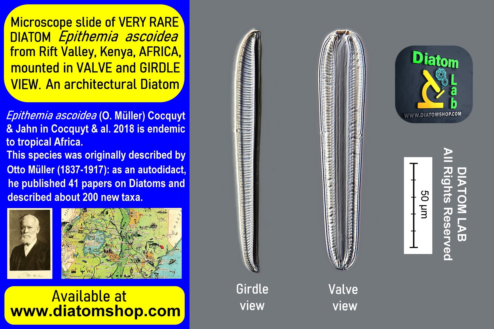
Microscope slide of 2 Very Rare African Diatoms Epithemia ascoidea from Rift Valley, Kenya, mounted in Valve and Girdle view! A very architectural Diatom! Mounted in the excellent Diatom Cubed mountant (refractive index > 1,7)
99.00 €
Add
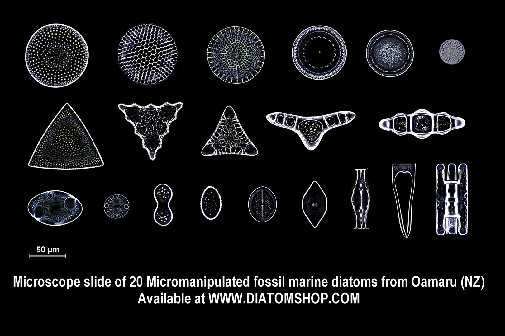
Microscope slide of 20 Selected mixed fossil marine diatoms from OAMARU, NZ (Upper Eocene Epoch: from 37.2 +/- 0.1 To 33.9+/- 0.1 million years ago), with Species list printed on the label. Micromanipulated in parallel lines (without duplicates) and mounted in the excellent Diatom Cubed mountant (refractive index > 1,7). See example in dark field illumination on the side: this sample contains many more species!
249.00 €
Add
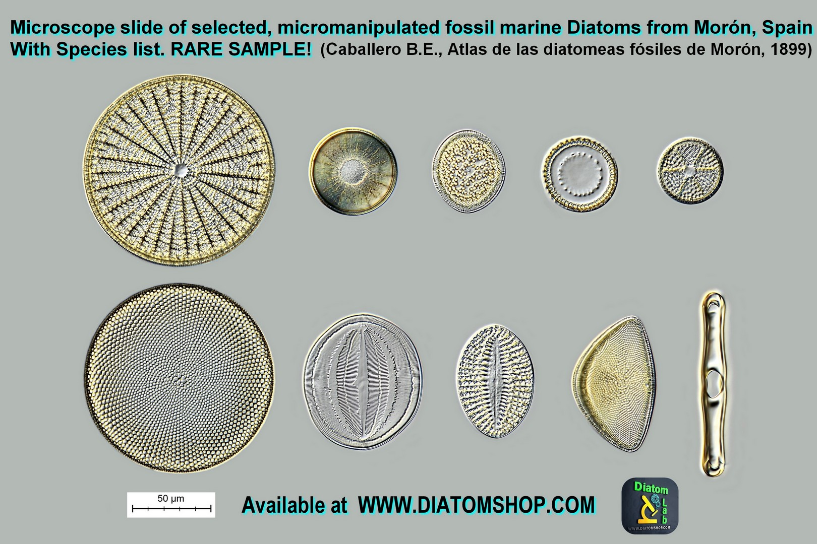
Microscope slide of 10 selected micromanipulated fossil marine diatoms from MORON, SPAIN, with Species List printed on the label ( RARE SAMPLE!). In this laboratory there's the same very rare rock that Ernesto Bellido Caballero used to publish his "Atlas de las diatomeas fósiles de Morón" in 1899. This sample has been carefully cleaned at Diatom Lab and contains about 170 diatom species, including the rare Asteromphalus moronensis (Greville) A.W.F.Schmid; Biddulphia moronensis (Greville) T.V.Desikachary; Euodia gibba var. moronensis Tempère & Peragallo, 1909; Grammatophora moronensis Greville 1863; Actinocyclus hispanicus F.Azpeitia Moros ; Cocconeis moronensis A.W.F.Schmidt; Coscinodiscus ovalis Roper 1858. Micromanipulated in two rows, without duplicates, mounted in the excellent Diatom Cubed mountant (refractive index > 1,7). See example in DIC on the side: this sample contains many more species!
149.00 €
Add
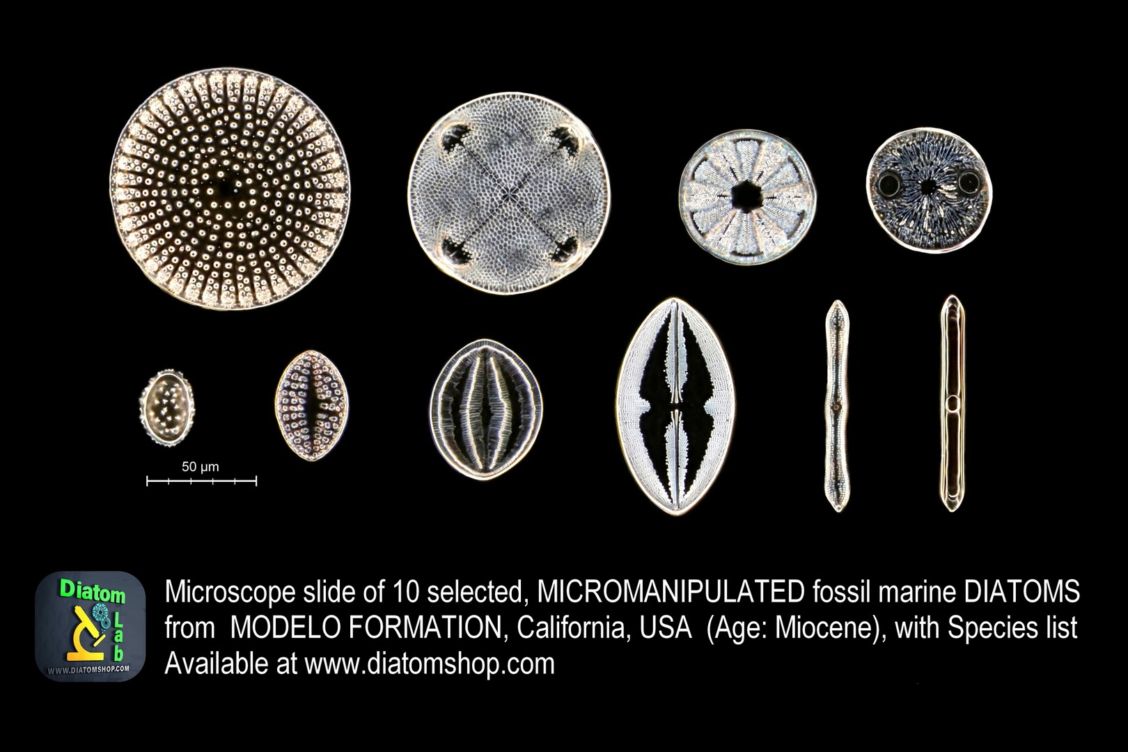
Microscope slide of 10 selected Micromanipulated fossil marine Diatoms from MODELO FORMATION, California, USA, with Species list printed on the label. Micromanipulated in parallel lines (without duplicates) and mounted in the excellent Diatom Cubed mountant (refractive index > 1,7). See example in dark field illumination on the side: this sample contains many more species!
149.00 €
Add
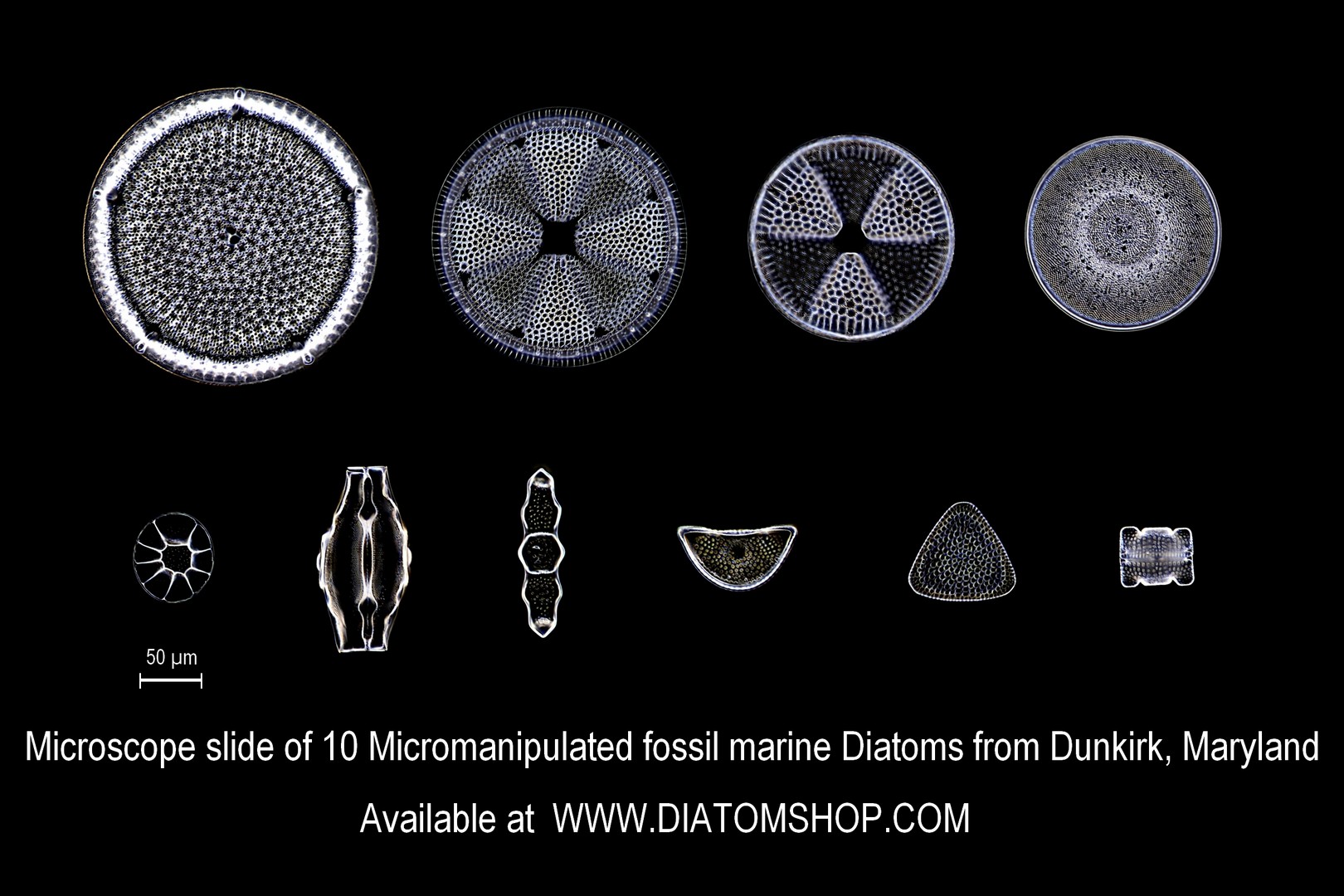
Microscope slide of 10 Selected mixed fossil marine diatoms from DUNKIRK, Maryland, USA (Miocene) with Species list printed on the label. Micromanipulated in parallel lines (without duplicates) and mounted in the excellent Diatom Cubed mountant (refractive index > 1,7). See example in dark field on the side: this sample contains many more species
149.00 €
Add
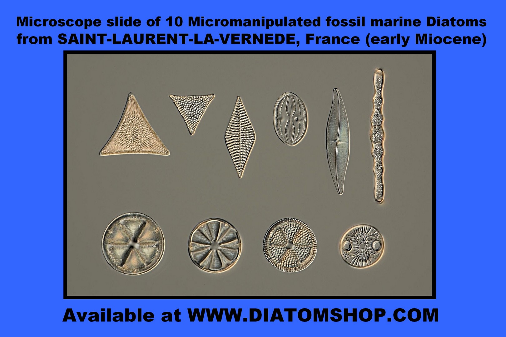
Microscope slide of 10 Selected mixed fossil marine diatoms from SAINT-LAURENT-LA-VERNEDE, France (early Miocene), with Species list printed on the label. See example in DIC on the side: this sample contains many more species! Diatoms are micromanipulated in parallel lines (without duplicates), mounted in the excellent Diatom Cubed mountant (refractive index > 1,7)
149.00 €
Add
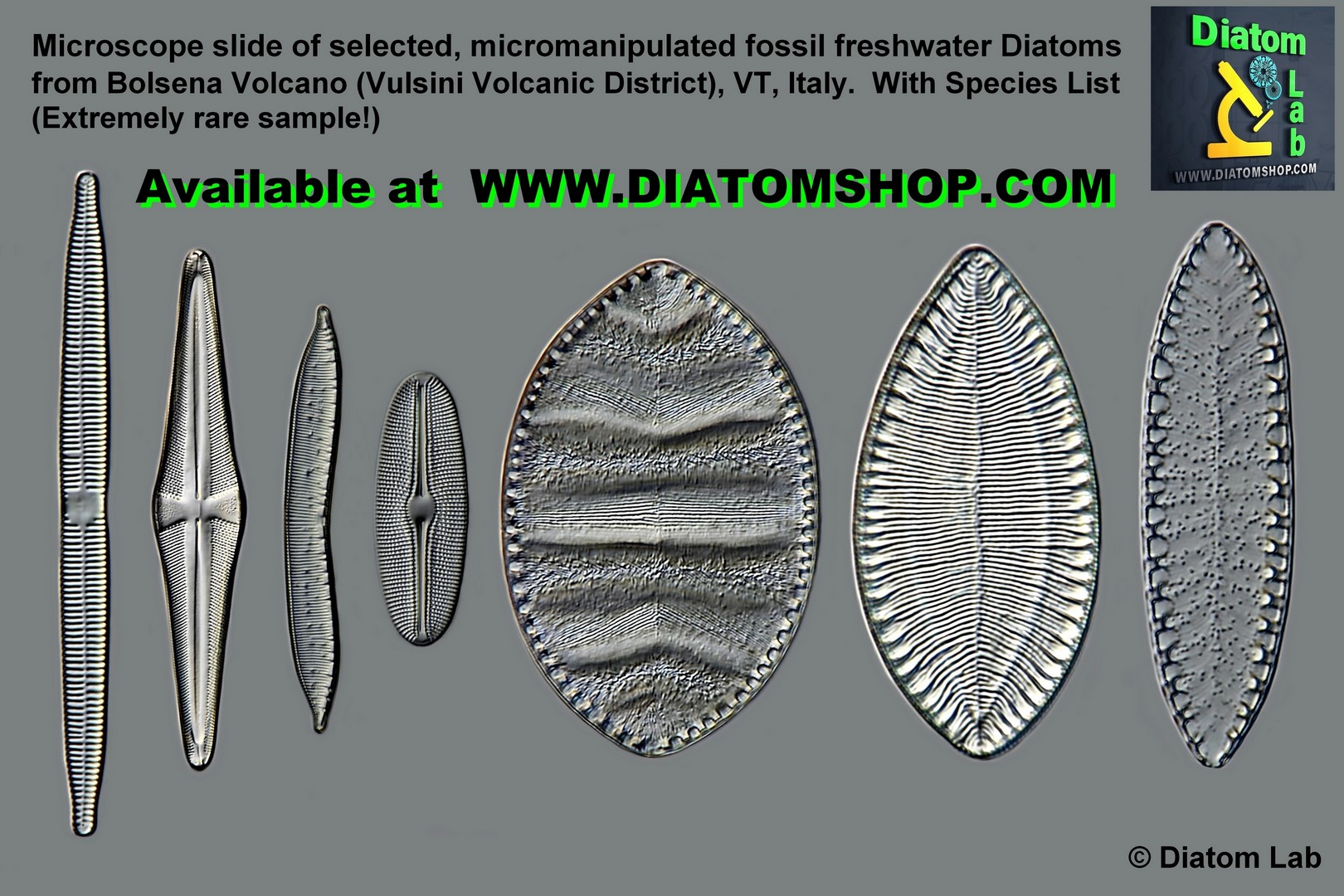
Microscope slide of 7 selected micromanipulated fossil freshwater diatoms from BOLSENA VOLCANO (Vulsini Volcanic District), VT, Italy, with Species List printed on the label (EXTREMELY RARE SAMPLE!). Vulsini Volcanic District is a caldera complex that created some Pleistocene lakes! Micromanipulated in a row, without duplicates, mounted in the excellent Diatom Cubed mountant (refractive index > 1,7). See example in DIC on the side: this sample contains many more species!
149.00 €
Add
Microscope slide of 10 Selected mixed fossil marine diatoms from SANTA MONICA, California, USA with Species list printed on the label. Micromanipulated in parallel lines (without duplicates) and mounted in the excellent Diatom Cubed mountant (refractive index > 1,7).This sample contains really many species!
169.00 €
Add
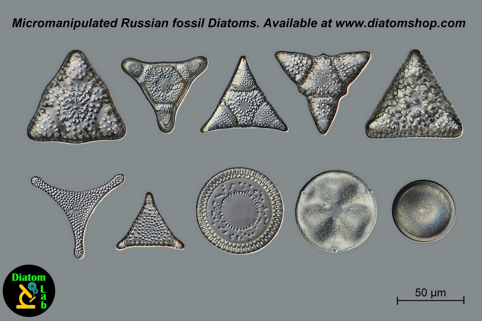
Microscope slide of 10 Selected mixed fossil marine diatoms from Inzensky District, RUSSIA (Cretaceous: from 145.5 to 65.5 million years ago, when dinosaurs - such as Tyrannosaurus - where numerous!!!), with Species list printed on the label. Micromanipulated in parallel lines (without duplicates) and mounted in the excellent Diatom Cubed mountant (refractive index > 1,7). Look at the examples on the side: this Russian sample contains many more species!
169.00 €
Add
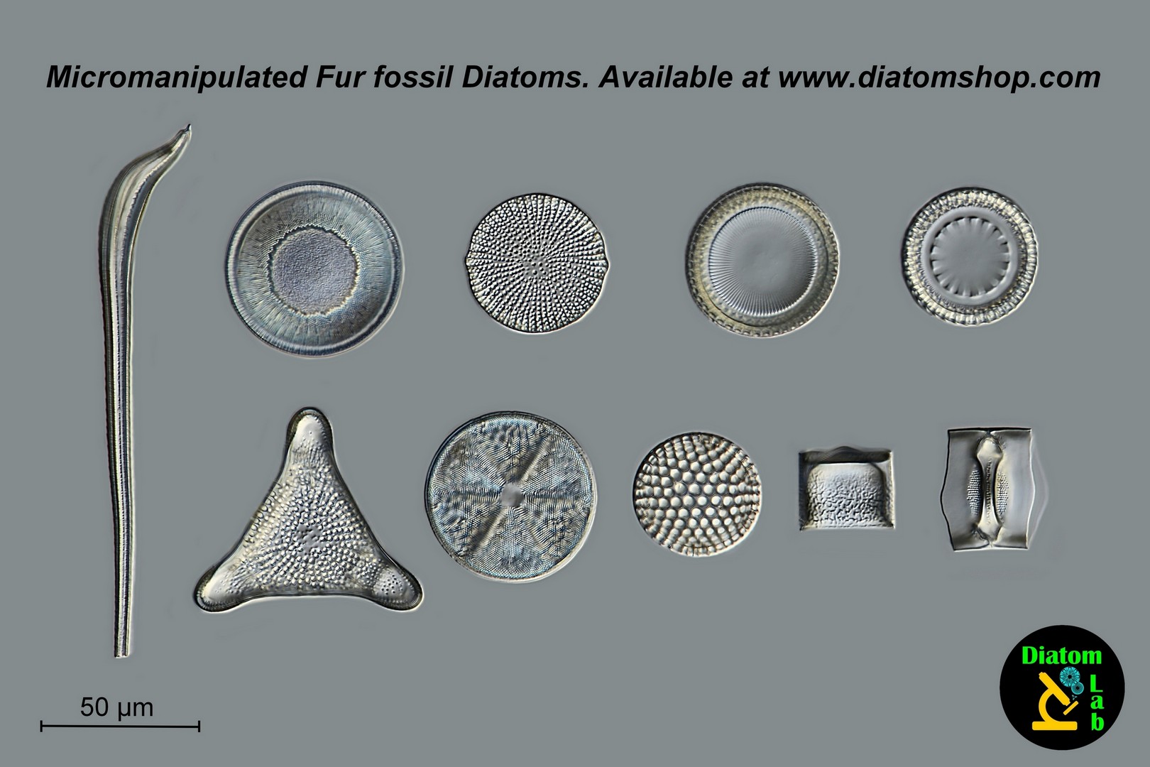
Microscope slide of 10 Selected mixed fossil marine diatoms from FUR ISLAND, Denmark (Paleogene: from 65.5 million to 23.0 million years ago) with Species list printed on the label. Micromanipulated in parallel lines (without duplicates) and mounted in the excellent Diatom Cubed mountant (refractive index > 1,7). See example in DIC on the side: this sample contains many more species
149.00 €
Add
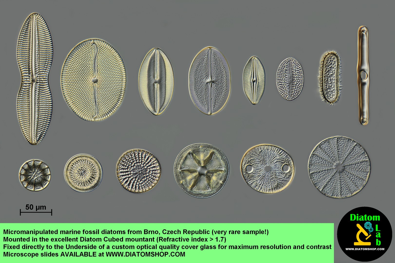
Microscope slide of 14 Micromanipulated fossil diatoms from BRNO, Czech Republic (Age: Langhian) with Species List printed on the label (EXTREMELY RARE SAMPLE!) Micromanipulated rows, without duplicates, mounted in the excellent Diatom Cubed mountant (refractive index > 1,7). Look at the example on the side, in DIC: this sample contains many more species
199.00 €
Add
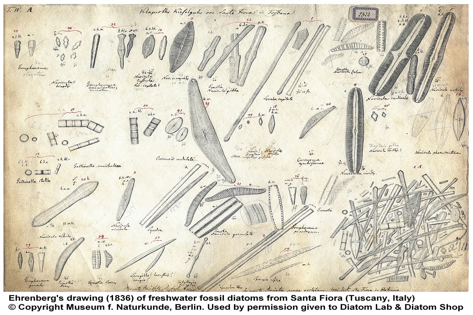
Microscope slide of 6 Selected micromanipulated fossil freshwater diatoms from SANTA FIORA, Tuscany, Italy (Miocene), with Species List printed on the label (EXTREMELY RARE SAMPLE!): the mines have been all walled up - destroyed around 1970!), initially studied by EHRENBERG in the 19th century: see Ehrenberg's drawings (1836) below! Micromanipulated in a row, without duplicates, mounted in the excellent Diatom Cubed mountant (refractive index > 1,7). Freshwater fossil diatoms from Santa Fiora have been initially described in: Ehrenberg Christian Gottfried, Die infusionsthierchen als vollkommene organismen. Ein blick in das tiefere organische leben der natur, Leipzig,L. Voss, 1838
149.00 €
Add
Microscope slide of 10 Selected mixe fossil marine diatoms from MORS ISLAND, Denmark (Paleogene: from 65.5 million to 23.0 million years ago) with Species list printed on the label. Micromanipulated in parallel lines (without duplicates) and mounted in the excellent Diatom Cubed mountant (refractive index > 1,7).
149.00 €
Add
Microscope slide of 10 selected mixed (recent - fossil - marine - freshwater) diatoms with SPECIES LIST printed on the label. Micromanipulated in parallel lines (without duplicates), mounted in the excellent Diatom Cubed mountant (refractive index > 1,7)
169.00 €
Add
Microscope slide of 16 Selected mixed (recent - fossil - marine - freshwater) diatoms with SPECIES LIST printed on the label. Micromanipulated in parallel lines (without duplicates), mounted in the excellent Diatom Cubed mountant (refractive index > 1,7)
199.00 €
Add
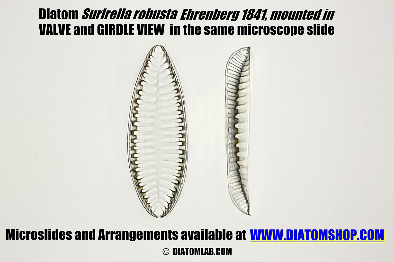
Diatoms with scientific names mounted both in VALVE and GIRDLE VIEW (in the same microscope slide), in this way you can fully appreciate all diatom structures! Please write us the species you prefer: Tetracyclus celatom Okuno (rare!) in valve and girdle view; Stephanogonia danica Grunow (rare!) in valve and girdle view; Anomoeoneis polygramma (Ehrenberg) Cleve 1895 in valve and girdle view; Epithemia turgida (Ehrenberg) Kützing 1844 in valve and girdle view. Mounted in the excellent Diatom Cubed mountant (refractive index > 1,7). Look at the example on the side
99.00 €
Add
Microscope slide of selected mixed Diatoms arranged in MEDIUM CIRCLE (approx. diameter 350 µm), mounted in the excellent Diatom Cubed mountant (refractive index > 1,7)
290.00 €
Add
Microscope slide of selected mixed fossil marine Diatoms from Oamaru (NZ) arranged in MEDIUM CIRCLE (approx. diameter 350 µm), mounted in the excellent Diatom Cubed mountant (refractive index > 1,7)
290.00 €
Add
Microscope slide of selected mixed Diatoms arranged in BIG CIRCLE (approx. diameter 430 µm), mounted in the excellent Diatom Cubed mountant (refractive index > 1,7). Look at the example on the side
490.00 €
Add
Microscope slide of 5 selected, micromanipulated PINNULARIA species, with Species list printed on the label: 1) Pinnularia cardinalis (Ehrenberg) W.Smith; 2) Pinnularia major (Kützing) Rabenhorst; 3) Pinnularia dactylus var. dariana (A.Schmidt) Cleve; 4) Pinnularia nobilis (Ehrenberg) Ehrenberg; 5) Pinnularia viridis (Nitzsch) Ehrenberg. Mounted in the excellent Diatom Cubed mountant (refractive index > 1,7)
199.00 €
Add
Microscope slide of 6 selected, micromanipulated SURIRELLA species, with Species list printed on the label: 1) Surirella spiralis Kützing; 2) Surirella helvetica Brun; 3) Surirella ovalis Brébisson; 4) Surirella fuellebornii f. subconstricta O.Müller; 5) Surirella utahensis (Grunow) Hanna & Grant; 6) Surirella striatula Turpin. Mounted in the excellent Diatom Cubed mountant (refractive index > 1,7)
249.00 €
Add
Set of 2 Microscope slides that help to VERIFY THE IMPORTANCE OF REFRACTIVE INDEX: one microscope slide contains the micromanipulated diatom Stauroneis phoenicenteron (Nitzsch) Ehrenberg mounted in DIATOM³ (Diatom Cubed) mountant (refractive index > 1.7), the other microscope slide contains the micromanipulated diatom Stauroneis phoenicenteron (Nitzsch) Ehrenberg mounted in Canada balsam (refractive index 1.52, near to that of ordinary glass)
199.00 €
Add
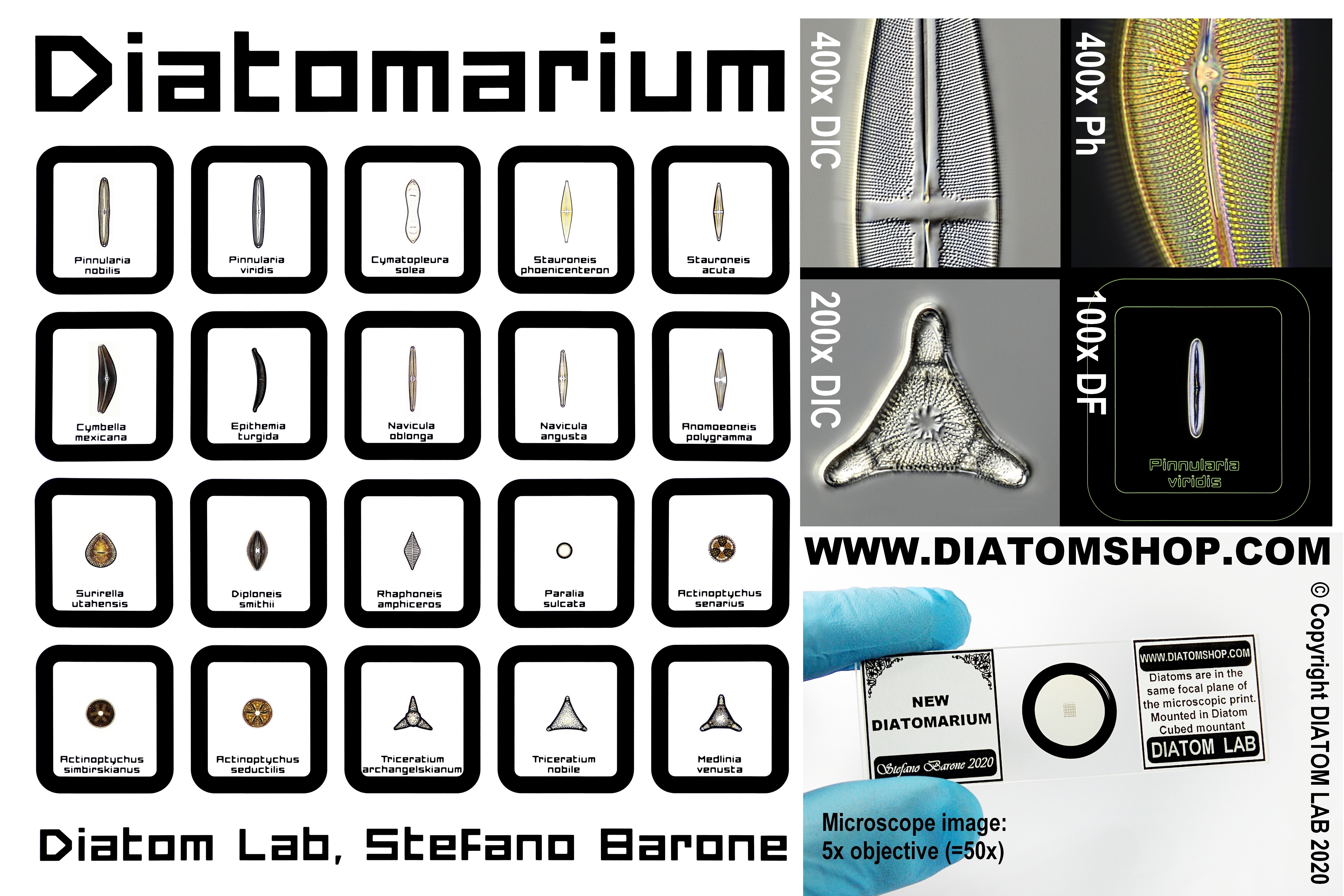
NEW DIATOMARIUMS: these are the first diatom microscope slides in the world made with this innovative, expensive and accurate technology! Microscope slides of micromanipulated diatom specimens mounted inside a revolutionary new grid with engraved scientific names or numbers. The letters of the species name are 16 µm high and each square internally measures 300 µm x 300 µm. The diatom array and the product & company names sit comfortably in the field of view of every 5x objective.
The New Diatomarium with engraved Scientific names is a COMPENDIUM of the Diatom world: in fact the New Diatomarium includes Recent Marine - Recent Freswater -Fossil Marine - Fossil Freshwater, Centric and Pennate species!
FOR the HIGH RESOLUTION IMAGE CLICK here
FOR the HIGH RESOLUTION IMAGE CLICK here
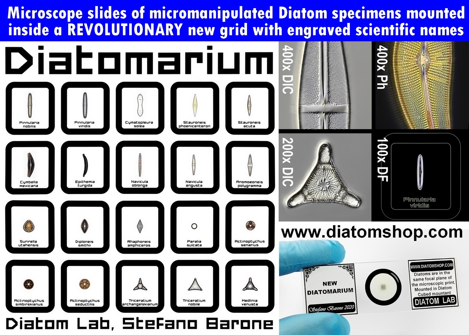
UNIQUE PRODUCT! Microscope slide of 20 micromanipulated diatom specimens mounted inside a revolutionary new GRID with engraved scientific names See images on the side.
Specimens belonging to Recent Marine samples: Actinoptychus senarius, Paralia sulcata,Rhaphoneis amphiceros
Specimens belonging to Fossil Marine samples: Actinoptychus simbirskianus, Actinoptychus seductilis, Triceratium archangelskianum , Triceratium nobile, Medlinia venusta
Specimens belonging to Freshwater Recent samples: Pinnularia nobilis, Stauroneis phoenicenteron, Pinnularia viridis, Cymatopleura solea, Navicula oblonga
Specimens belonging to Freshwater Fossil samples: Stauroneis acuta, Epithemia turgida,Navicula angusta, Cymbella mexicana, Anomoeoneis polygramma, Surirella utahensis, Diploneis smithii
399.00 €
Add
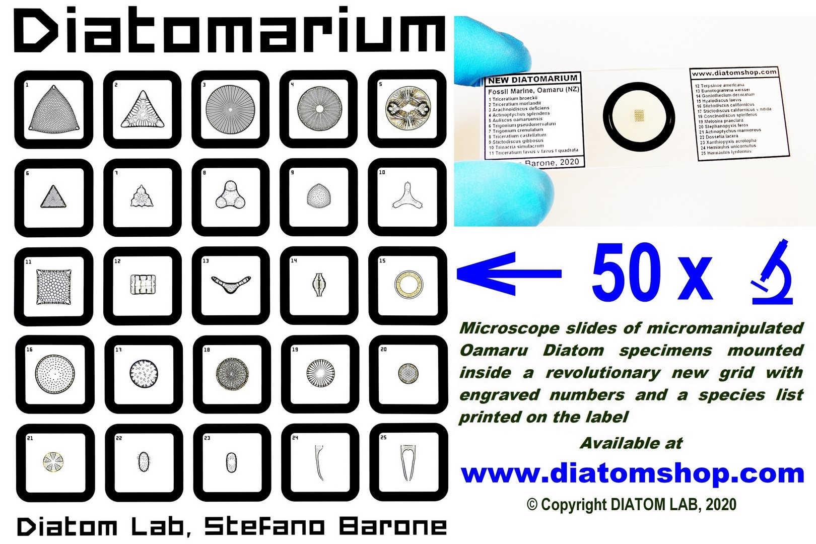
UNIQUE PRODUCT! Microscope slides of 25 Micromanipulated OAMARU Diatom specimens mounted inside a revolutionary new GRID with engraved numbers and a species list printed on the label. The rare Oamaru (marine fossil) Diatoms are among the most beautiful in the world, with more than 600 taxa! Each New Diatomarium is unique as contains different Oamaru Diatoms: see example on the side
399.00 €
Add
SINGLE DIATOM MOUNTS (MICROSCOPE SLIDES OF ONE SELECTED, MICROMANIPULATED DIATOM). All micromanipulated Diatoms are guaranteed to be fixed directly to the UNDERSIDE of the custom, optical quality cover glass (and not, as is common, on the microscope slide) for maximum resolution and contrast! The reason for this is that: microscope objective performance drops quickly and noticeably as the specimen distance from the cover glass increases. Microscope objective lenses are designed to be optimally corrected for objects located immediately below the coverslip!
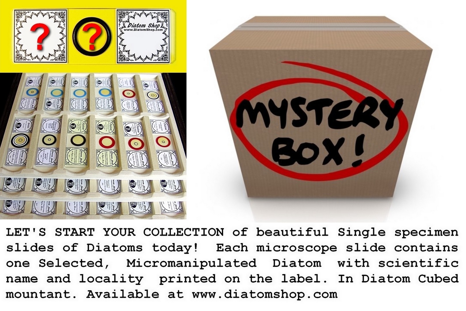
Let's start your COLLECTION of beautiful Single specimen slides of Diatoms today! Each MYSTERY microscope slide contains one Selected, Micromanipulated Diatom with scientific name and locality printed on the label. In Diatom Cubed mountant. The price is for one MYSTERY microscope slide, and you can select the desired quantity of purchase
39.00 €
Add
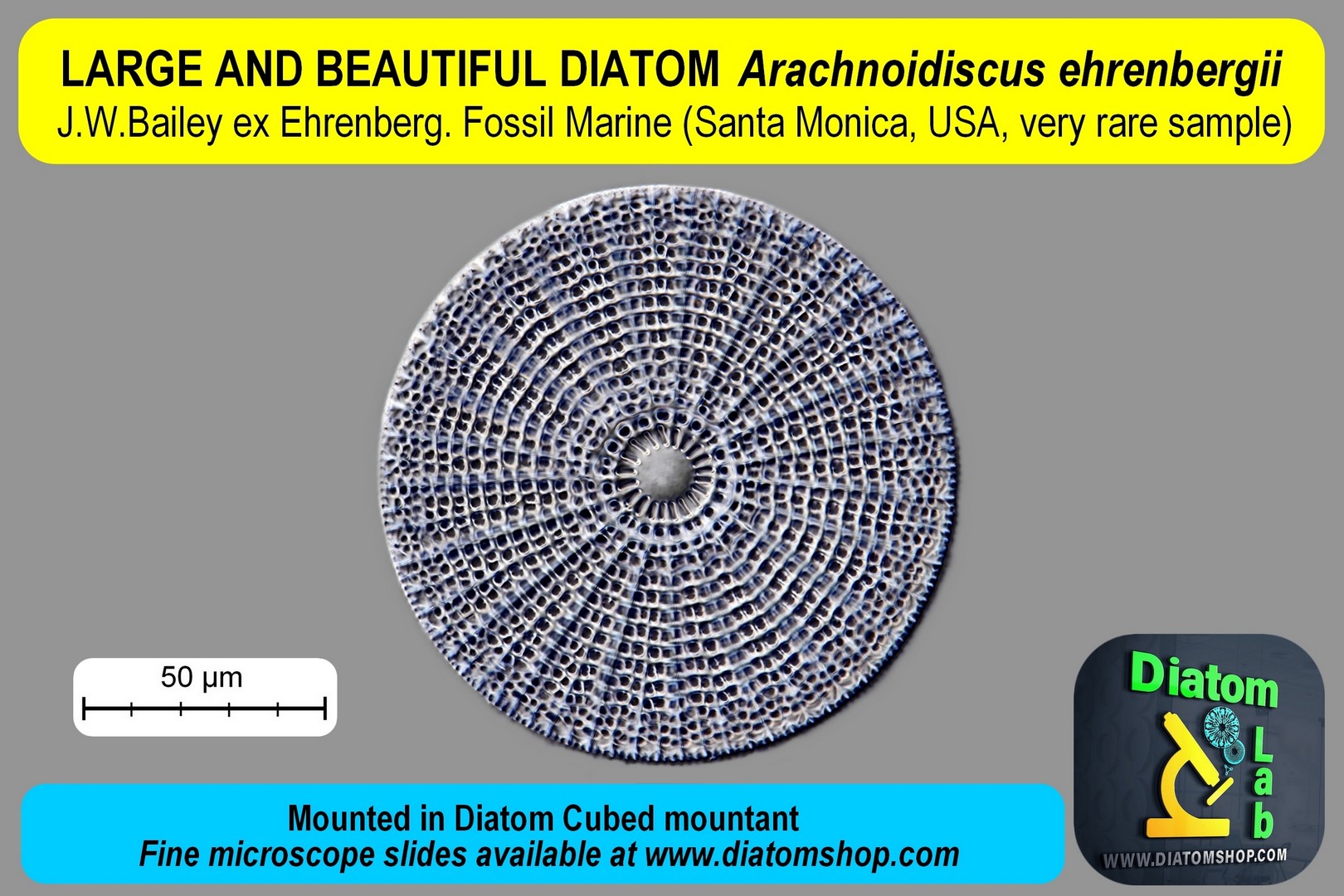
Single diatom mount. Microscope slide of Arachnoidiscus ehrenbergii J.W.Bailey ex Ehrenberg. Mounted in Diatom Cubed mountant
69.00 €
Add
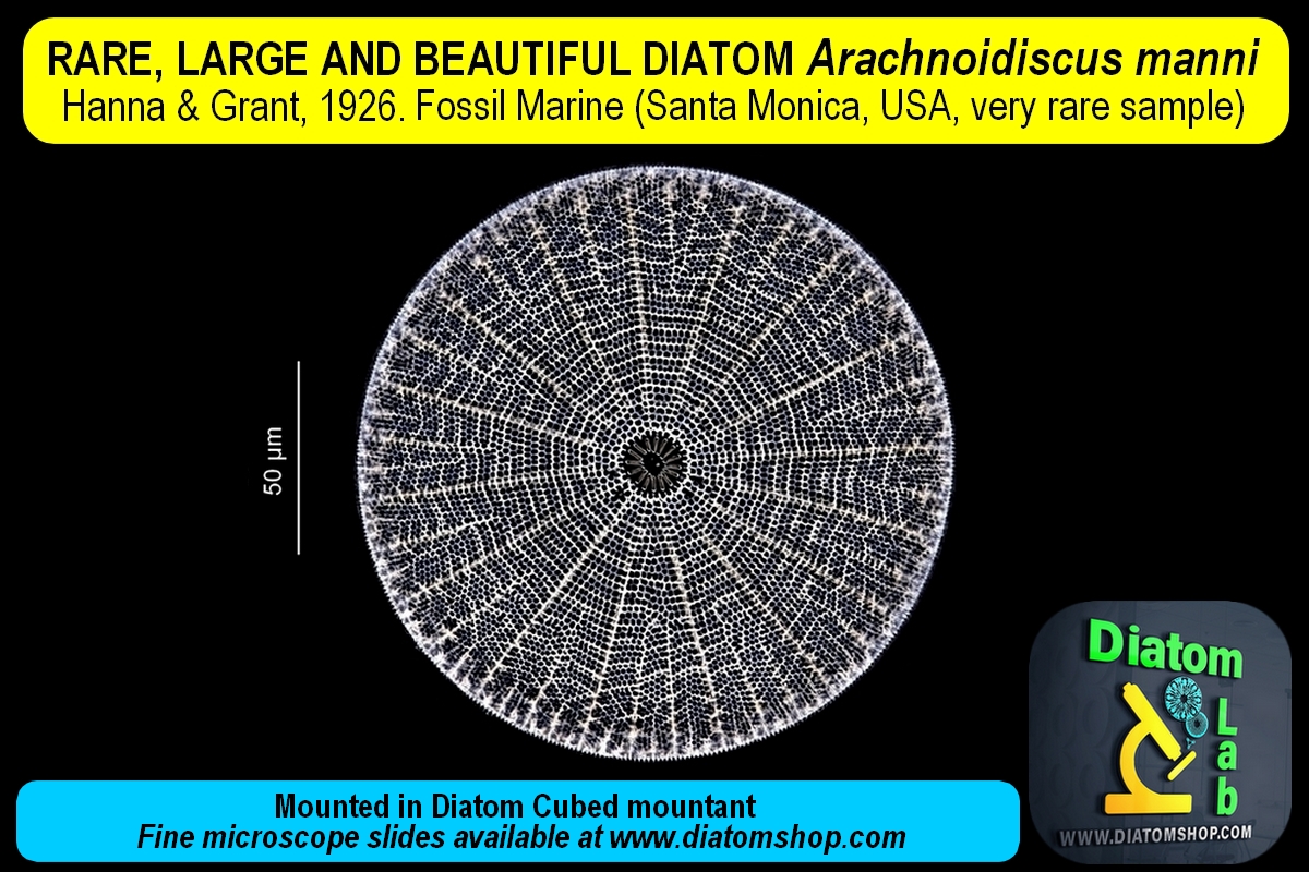
Single diatom mount. Microscope slide of Arachnoidiscus manni Hanna & Grant, 1926. Mounted in Diatom Cubed mountant
69.00 €
Add

Microscope slide of thick, large Diatom, excellent for making 3D MODELS of diatoms (images and movies) by means of a software (such as Helicon Focus, Zerene Stacker, Combine ZP and Adobe Photoshop CS4 / CS5 / CS6). Mounted in the excellent Diatom Cubed mountant (refractive index > 1,7). You can download the publication "Advanced photomicrography: construction of a 3D Diatom model saved as an image or movie" by the page about Publications
69.00 €
Add
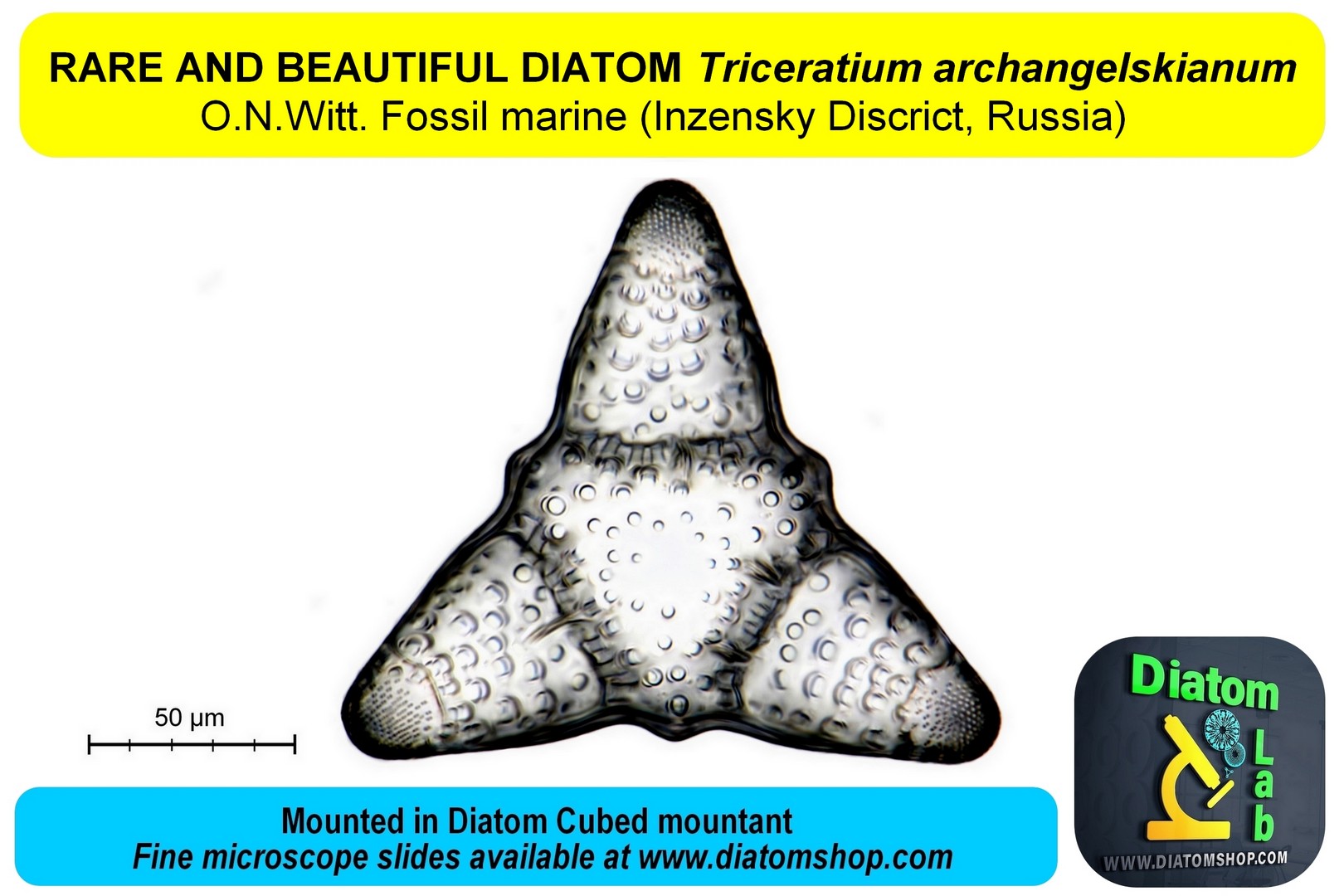
Single diatom mount. Microscope slide of Triceratium archangelskianum O.N.Witt. Mounted in Diatom Cubed mountant.
69.00 €
Add

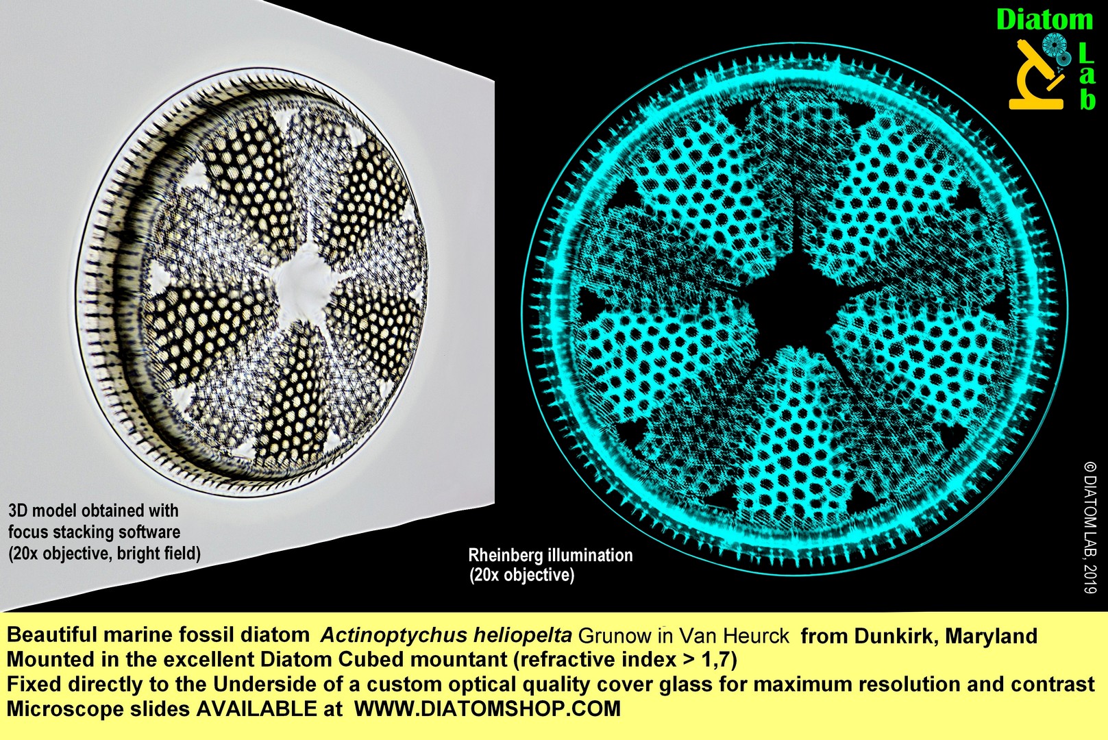
Single diatom mount. Microscope slide of Actinoptychus heliopelta Grunow in Van Heurck 1883 from Dunkirk, Maryland, USA. Mounted in Diatom Cubed mountant.
59.00 €
Add
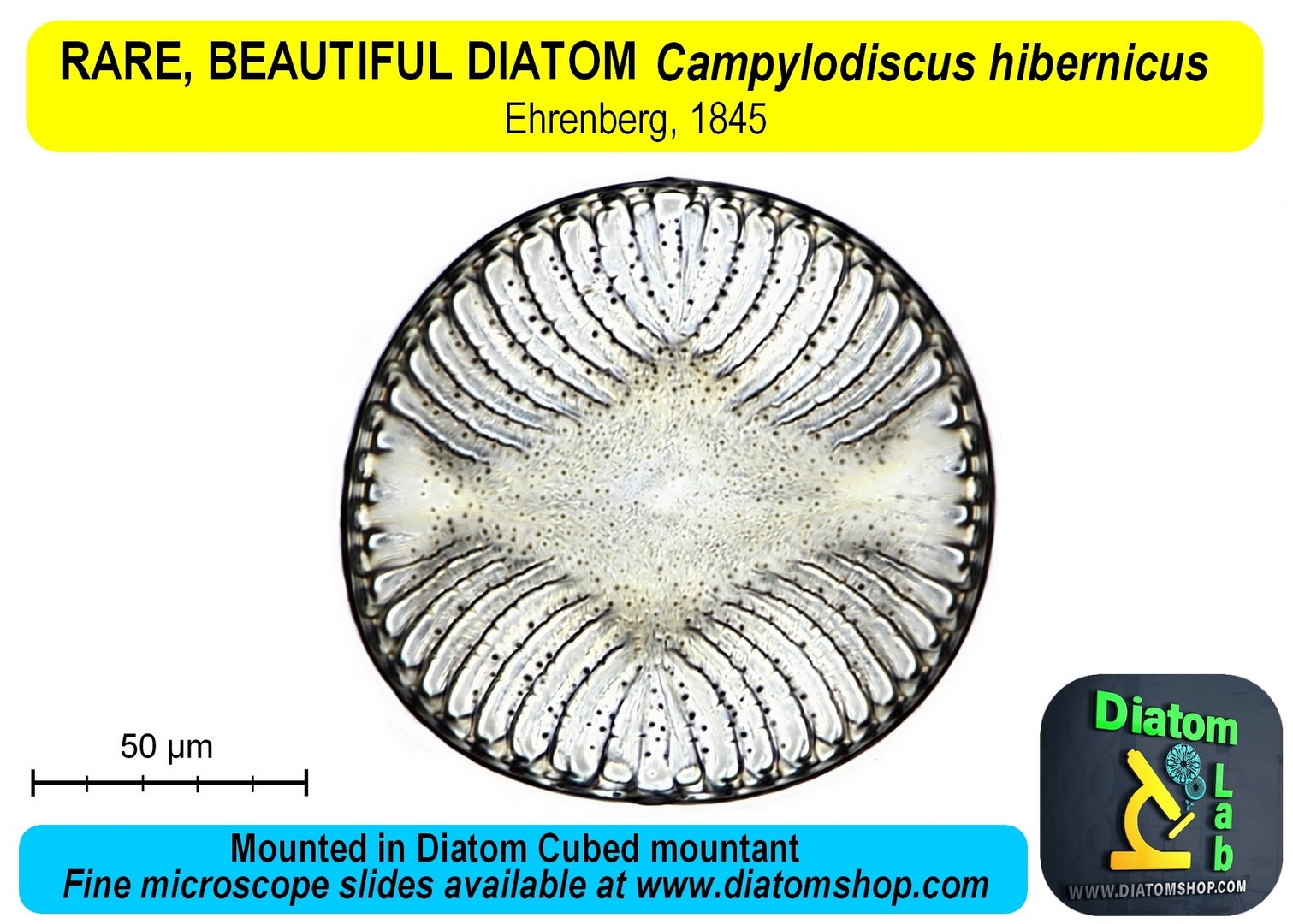
Single diatom mount. Microscope slide of extremely rare Campylodiscus hibernicus Ehrenberg. Mounted in Diatom Cubed mountant
69.00 €
Add
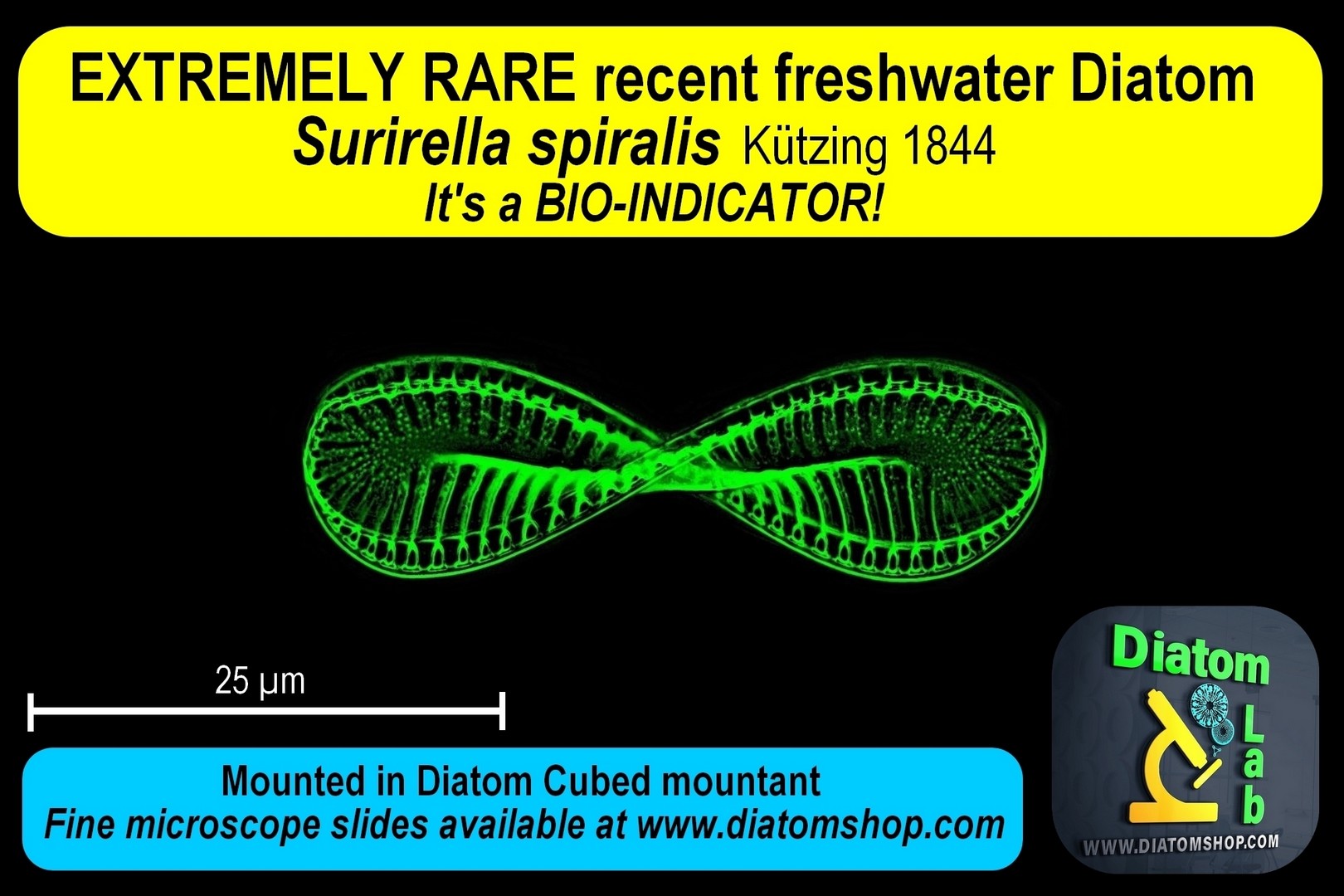
Single diatom mount. Microscope slide of Extremely rare Surirella spiralis Kützing 1844 (bioindicator). Mounted in Diatom Cubed mountant
99.00 €
Add
MICROSCOPE SLIDES OF MICROMANIPULATED DIATOMS FORMING WORDS, INITIALS OR LETTERS! Click this link for a bigger image. A unique gift for you or your loved ones! You can ask for your customized arrangement! (Science, Art, Technology & Innovation by Diatom Lab). Only circular diatoms of the same species - Paralia sulcata (Ehrenberg) Cleve - are selected and perfectly micromanipulated! Each diatom has a diameter of about 30 µm and the letters are visible only under a light microscope! All micromanipulated Diatoms are guaranteed to be fixed directly to the UNDERSIDE of the custom optical quality cover glass (and not, as is common, on the microscope slide) for maximum resolution and contrast! The reason for this: microscope objective performance drops quickly noticeably as the specimen distance from the cover glass increases. Microscope objective lenses are designed to be optimally corrected for objects located immediately below the coverslip!
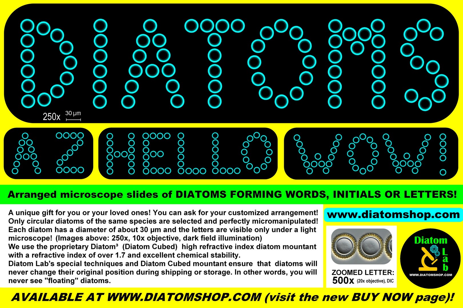
Arranged microscope slide of Diatoms forming ONE LETTER (AFTER PAYMENT, PLEASE WRITE AN EMAIL TO info@diatomshop.com AND SPECIFY THE REQUIRED LETTER). Only circular diatoms of the same species - Paralia sulcata (Ehrenberg) Cleve - are selected and perfectly micromanipulated. Mounted in the excellent Diatom Cubed mountant (refractive index > 1,7)
89.00 €
Add
Arranged microscope slide of Diatoms forming TWO LETTERS (AFTER PAYMENT, PLEASE WRITE AN EMAIL TO info@diatomshop.com AND SPECIFY THE REQUIRED LETTER). Only circular diatoms of the same species - Paralia sulcata (Ehrenberg) Cleve - are selected and perfectly micromanipulated. Mounted in the excellent Diatom Cubed mountant (refractive index > 1,7)
169.00 €
Add
Arranged microscope slide of Diatoms forming THREE LETTERS (AFTER PAYMENT, PLEASE WRITE AN EMAIL TO info@diatomshop.com AND SPECIFY THE REQUIRED LETTER). Only circular diatoms of the same species - Paralia sulcata (Ehrenberg) Cleve - are selected and perfectly micromanipulated. Mounted in the excellent Diatom Cubed mountant (refractive index > 1,7)
249.00 €
Add
Arranged microscope slide of Diatoms forming the word HELLO. Only circular diatoms of the same species - Paralia sulcata (Ehrenberg) Cleve - are selected and perfectly micromanipulated. Mounted in the excellent Diatom Cubed mountant (refractive index > 1,7)
299.00 €
Add
Arranged microscope slide of Diatoms forming the word DIATOMS. Only circular diatoms of the same species - Paralia sulcata (Ehrenberg) Cleve - are selected and perfectly micromanipulated. Mounted in the excellent Diatom Cubed mountant (refractive index > 1,7)
349.00 €
Add
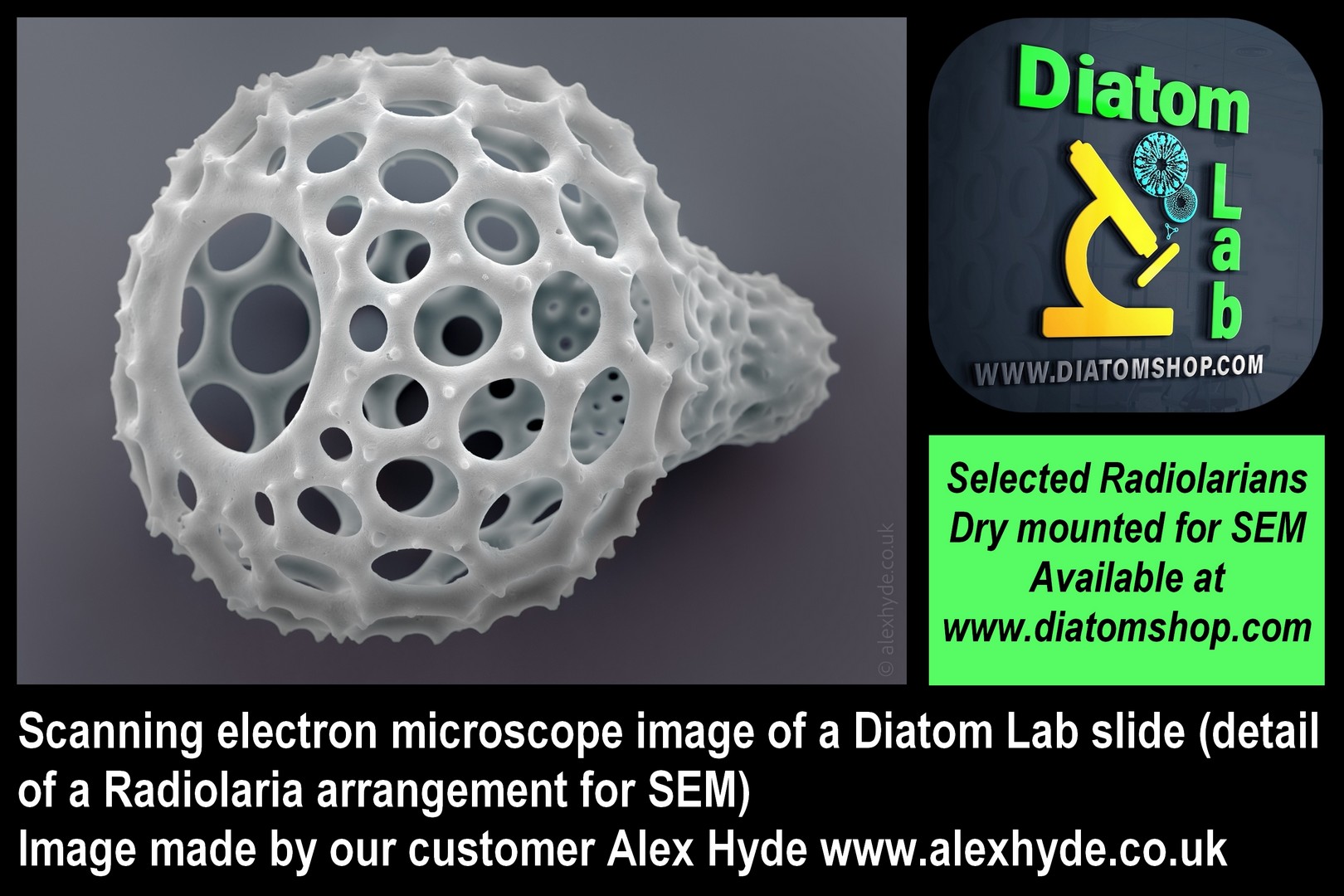
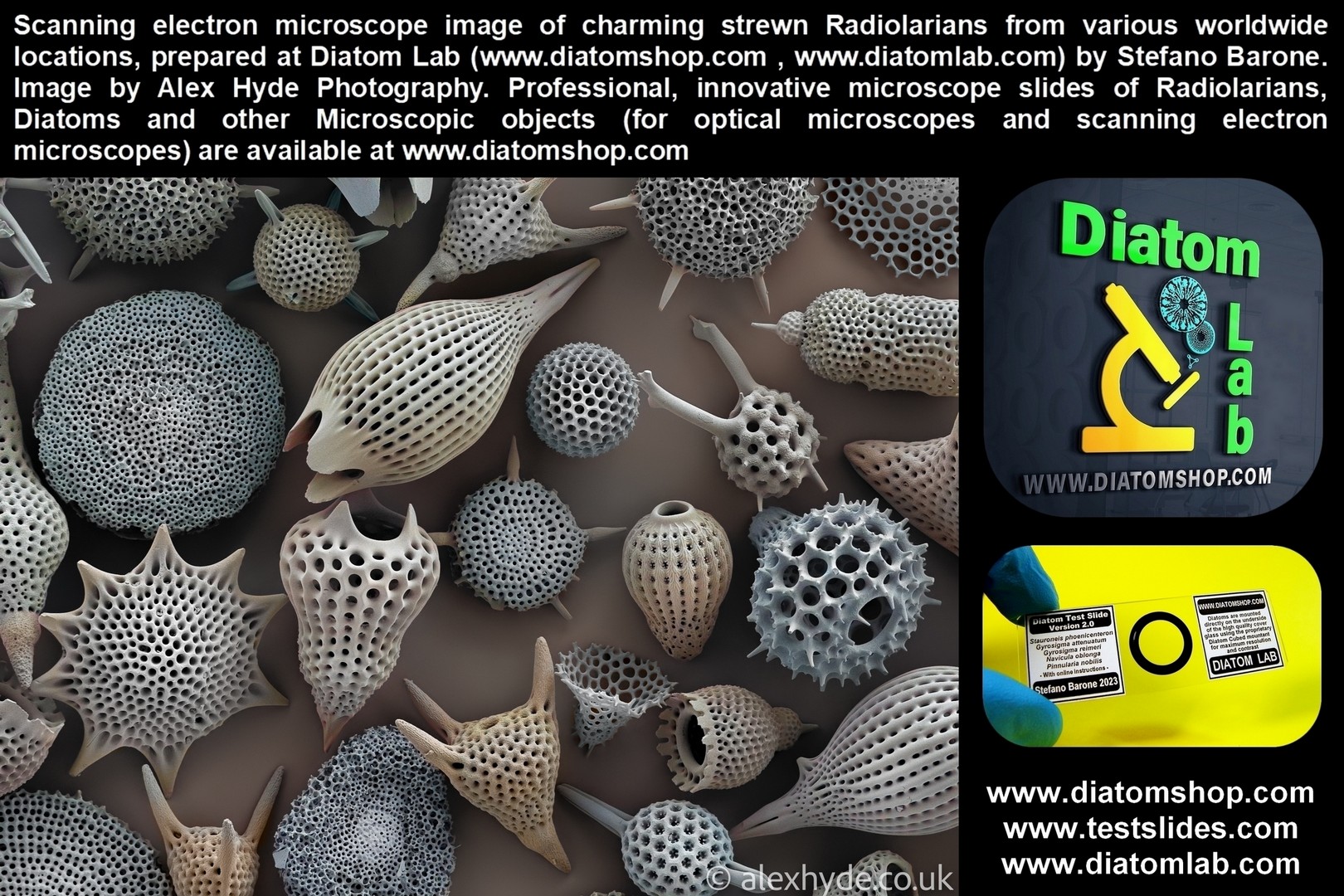
RADIOLARIANS: MICROSCOPE SLIDES. As in the case of micromanipulated Diatoms, all micromanipulated Radiolarians are guaranteed to be fixed directly to the UNDERSIDE of the custom optical quality cover glass (and not, as is common, on the microscope slide) for maximum resolution and contrast! The reason for this: microscope objective performance drops quickly noticeably as the specimen distance from the cover glass increases. Microscope objective lenses are designed to be optimally corrected for objects located immediately below the coverslip!
Microscope slide of 10 selected, micromanipulated Radiolarians from Barbados, Bath Cliff, RARE locality, without duplicates, arranged in parallel lines or in circle and mounted in Prime Mountant, excellent high refractive index mountant with refractive index > 1,7. See example in dark field illumination on the side (Described in the publication: Saunders, J.B., Bernoulli, D., Mueller-Merz, E., Oberhänsli, H., Perch-Nielsen, K., Riedel, W.R., Sanfilippo, A., and Torrini, R., Jr., 1984. Stratigraphy of the late middle Eocene to early Oligocene in the Bath Cliff section, Barbados, West Indies. Micropaleontology, 30:390−425).
159.00 €
Add
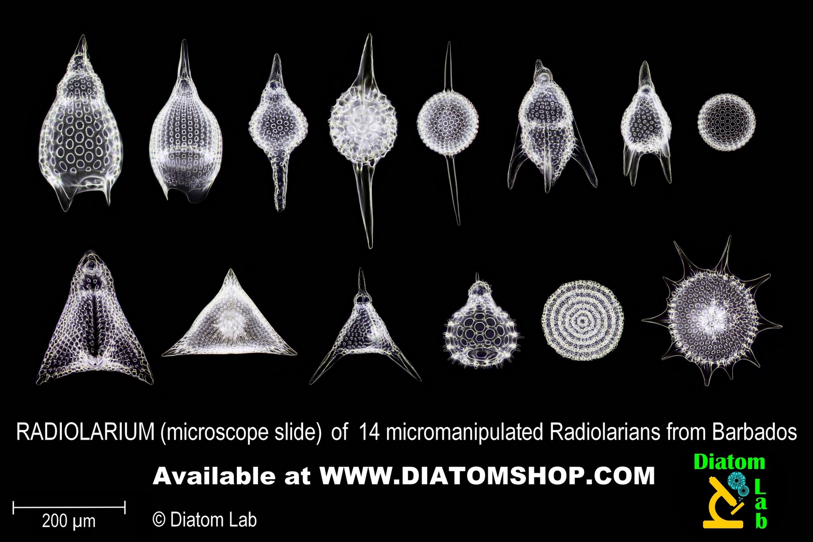
Microscope slide of 14 selected, micromanipulated Radiolarians from Barbados, Bath Cliff, RARE locality, without duplicates, arranged in parallel lines or in circle and mounted in Prime Mountant, excellent high refractive index mountant with refractive index > 1,7. See example in dark field illumination on the side (Described in the publication: Saunders, J.B., Bernoulli, D., Mueller-Merz, E., Oberhänsli, H., Perch-Nielsen, K., Riedel, W.R., Sanfilippo, A., and Torrini, R., Jr., 1984. Stratigraphy of the late middle Eocene to early Oligocene in the Bath Cliff section, Barbados, West Indies. Micropaleontology, 30:390−425)
199.00 €
Add
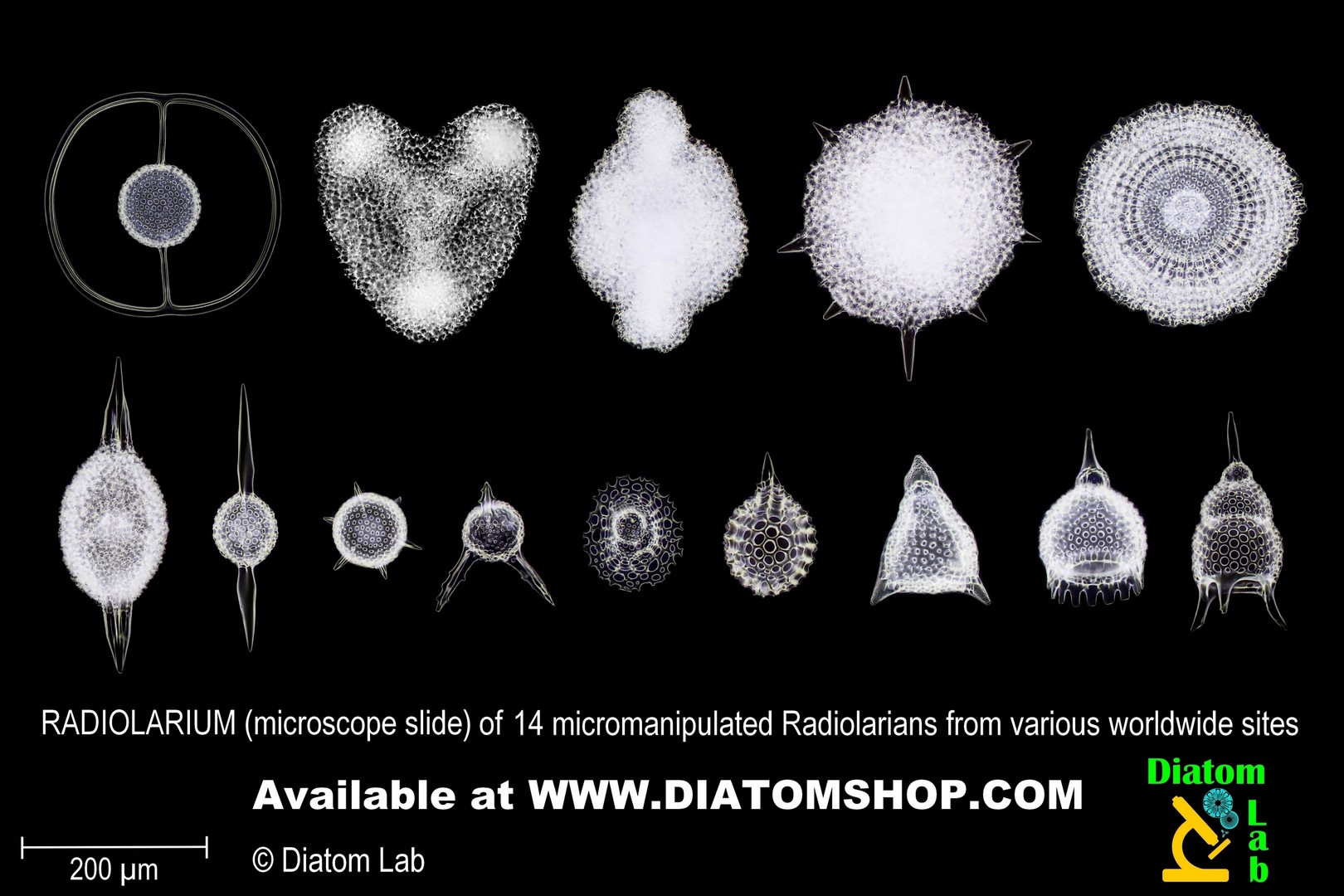
MEDIUM RADIOLARIUM © (microscope slide) of 14 selected and micromanipulated Radiolarians from various Worldwide sites, without duplicates, arranged in parallel lines or in circle and mounted in Prime Mountant, excellent high refractive index mountant with refractive index > 1,7. See example in dark field illumination on the side.
199.00 €
Add
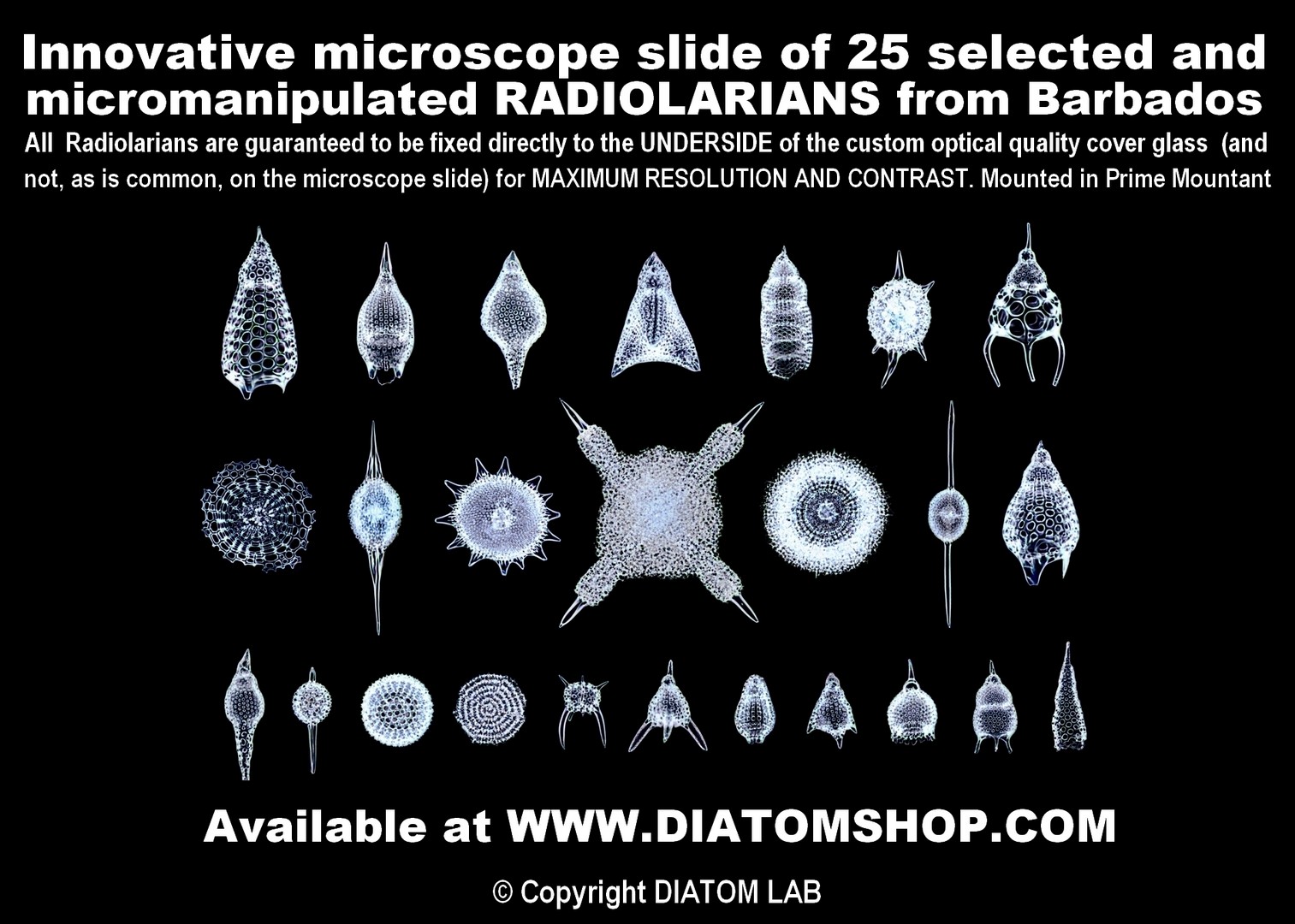
LARGE RADIOLARIUM © (microscope slide) of 25 selected, micromanipulated Radiolarians from Barbados, without duplicates, arranged in parallel lines or in circle and mounted in Prime Mountant, excellent high refractive index mountant with refractive index > 1,7. See example in dark field illumination on the side.
299.00 €
Add
LARGE RADIOLARIUM © (microscope slide) of 25 selected and micromanipulated Radiolarians from various Worldwide sites, without duplicates, arranged in parallel lines or in circle and mounted in Prime Mountant, excellent high refractive index mountant with refractive index > 1,7.
299.00 €
Add
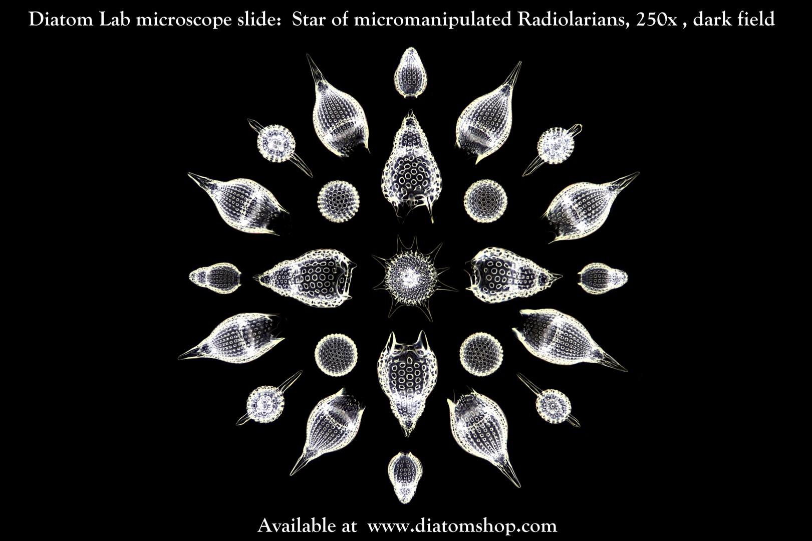
Star of selected, micromanipulated Radiolarians mounted in Prime Mountant, excellent high refractive index mountant with refractive index > 1,7. (See example on the side)
249.00 €
Add
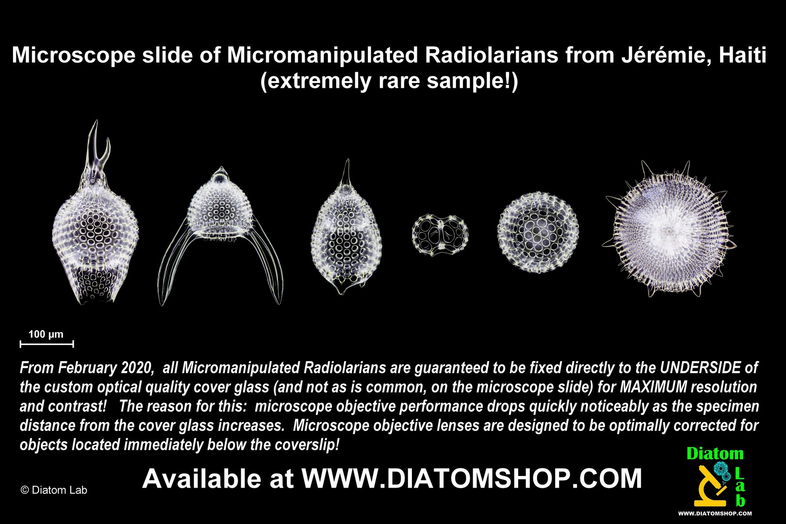
Microscope slide of 6 selected, micromanipulated Radiolarians from Jérémie, HAITI, Lower Miocene! EXTREMELY RARE SAMPLE: for Diatom Lab it was very difficult to collect several samples in Haiti, because of the current unstable and dangerous security situation in that country, located on the island of Hispaniola!
Without duplicates, arranged in line or in circle and mounted in Prime Mountant, excellent high refractive index mountant with refractive index > 1,7. See example in dark field illumination on the side
Without duplicates, arranged in line or in circle and mounted in Prime Mountant, excellent high refractive index mountant with refractive index > 1,7. See example in dark field illumination on the side
129.00 €
Add
Microscope slide of 10 selected, micromanipulated Radiolarians from Barbados, WITH SPECIES LIST PRINTED ON THE LABEL. Without duplicates, arranged in parallel lines or in circle and mounted in Prime Mountant, excellent high refractive index mountant with refractive index > 1,7.
179.00 €
Add
Microscope slide of 10 Micromanipulated Radiolarians from various Worldwide sites, with Species list
Microscope slide of 10 selected, micromanipulated Radiolarians from various Worldwide sites, WITH SPECIES LIST PRINTED ON THE LABEL. Without duplicates, arranged in parallel lines or in circle and mounted in Prime Mountant, excellent high refractive index mountant with refractive index > 1,7.
179.00 €
Add

Microscope slide of strewn Radiolarians from Valconca, RN, Italy. This rare fossil marine sample dates back to the Upper Miocene. The more you buy, the more chance you have of finding different species.
The image alongside shows selected and micromanipulated specimens, while in this microscope slide (“strew slide”) the specimens are not selected, they are scattered together with diatoms always belonging to the same sample.
The image alongside shows selected and micromanipulated specimens, while in this microscope slide (“strew slide”) the specimens are not selected, they are scattered together with diatoms always belonging to the same sample.
59.00 €
Add
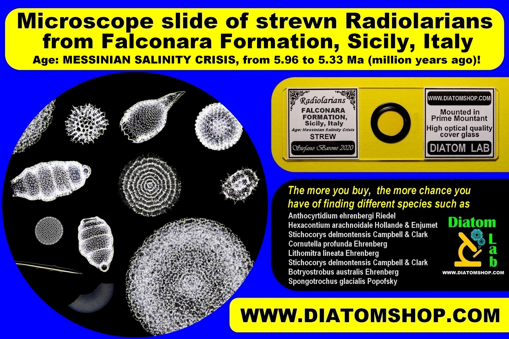
Microscope slide of strewn Radiolarians and Diatoms from FALCONARA FORMATION, Sicily, Italy. Age: MESSINIAN SALINITY CRISIS. VERY RARE!
EXTREMELY RARE Microscope slide of strewn Radiolarians from FALCONARA FORMATION. Age: MESSINIAN SALINITY CRISIS, from 5.96 to 5.33 Ma (million years ago)! Mounted in Prime Mountant, excellent high refractive index mountant with refractive index > 1,7. The more you buy, the more chance you have of finding different species!
EXTREMELY RARE Microscope slide of strewn Radiolarians from FALCONARA FORMATION. Age: MESSINIAN SALINITY CRISIS, from 5.96 to 5.33 Ma (million years ago)! Mounted in Prime Mountant, excellent high refractive index mountant with refractive index > 1,7. The more you buy, the more chance you have of finding different species!
59.00 €
Add
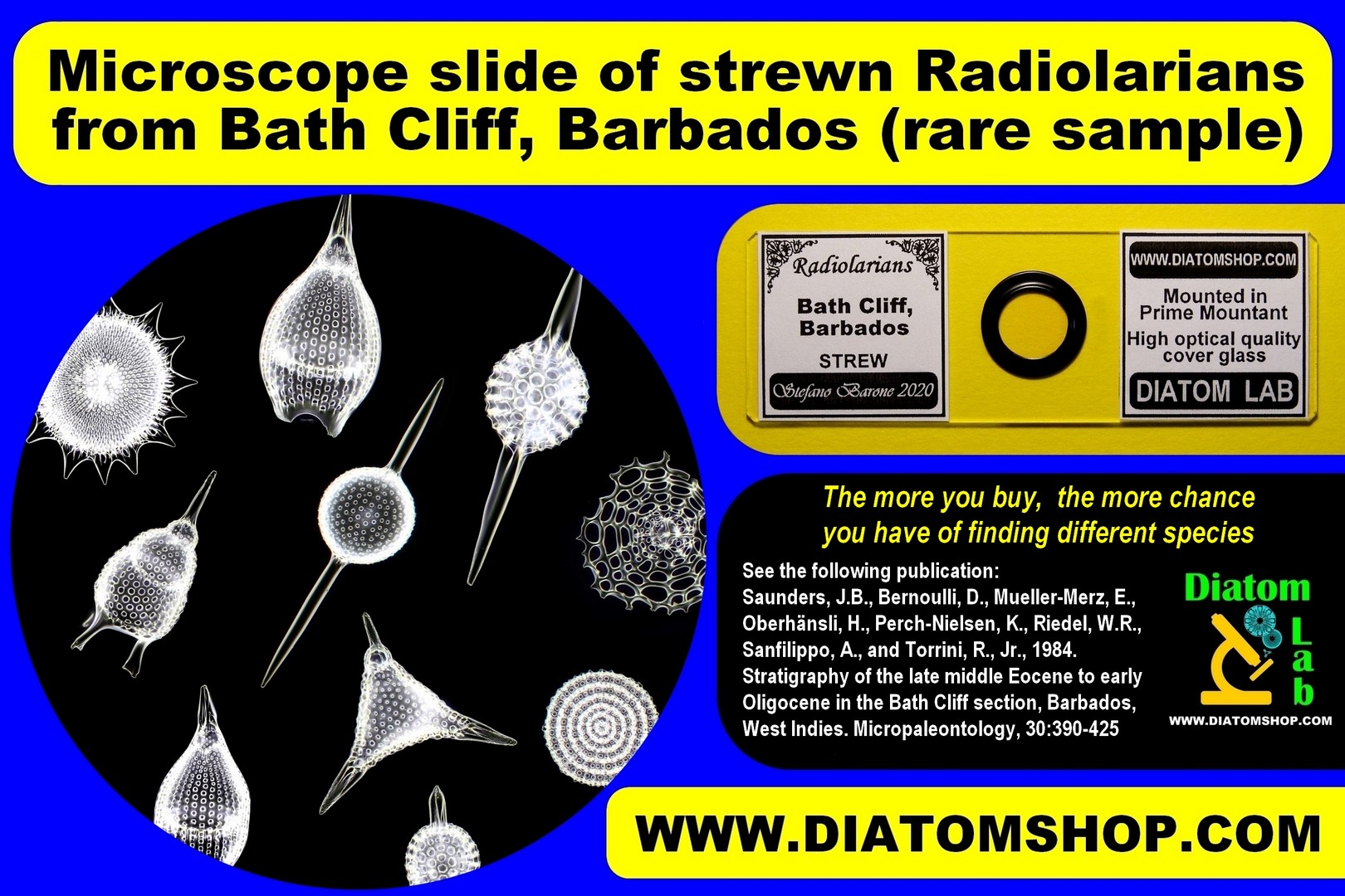
The more you buy, the more chance you have of finding different species!Microscope slide of strewn Radiolarians from Bath Cliff, RARE sample from Barbados, mounted in Prime Mountant, excellent high refractive index mountant with refractive index > 1,7. (Described in the publication: Saunders, J.B., Bernoulli, D., Mueller-Merz, E., Oberhänsli, H., Perch-Nielsen, K., Riedel, W.R., Sanfilippo, A., and Torrini, R., Jr., 1984. Stratigraphy of the late middle Eocene to early Oligocene in the Bath Cliff section, Barbados, West Indies. Micropaleontology, 30:390−425)
49.00 €
Add
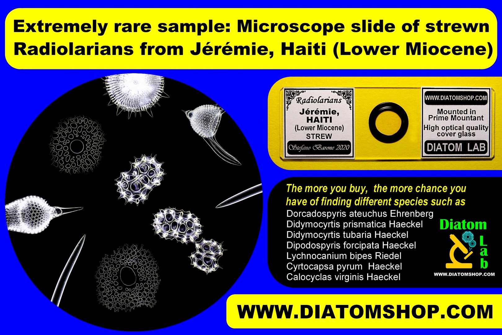
The more you buy, the more chance you have of finding different species! EXTREMELY RARE Microscope slide of strewn Radiolarians from Jérémie, HAITI (Lower Miocene). Mounted in Prime Mountant, excellent high refractive index mountant with refractive index > 1,7. For Diatom Lab it was very difficult to collect several samples in Haiti, because of the current unstable and dangerous security situation in that country, located on the island of Hispaniola!
See:
1) Riedel W. R., 1959. Oligocene and Lower Miocene Radiolaria in Tropical Pacific Sediments. Micropaleontology Vol. 5, No. 3, pp. 285-302
2)Truan y Luard, A. & Witt, O.N.,1888. Die Diatomaceen der Polycystinenkreide von Jeremie in Hayti, Westindien. Friedlander et Sohn, Berlin., 24 pp., 7 pls.
3) Möller, J. D., 1892. Polycystinen-Mergel von Jérémy, Haiti. (Verzeichnis der in den Lichtdrucktafeln Möller'scher Diatomaceen-Präparate enthaltenen Arten). Wedel (Holstein): Selbstverlag des Herausgebers, pp. 110-112, tafel 16
Our samples collected in Jérémie, HAITI contains several very rare Radiolarian species (and occasionally also several very rare Diatom species!), including:
Dorcadospyris ateuchus Ehrenberg, synonym: Ceratospyris ateuchus Ehrenberg 1873
Didymocyrtis prismatica Haeckel, synonym: Pipettella prismatica Haeckel, 1887
Didymocyrtis tubaria Haeckel, synonym: Pipettaria tubaria Haeckel, 1887
Dipodospyris forcipata Haeckel, 1887
Lychnocanium bipes Riedel, 1959
Cyrtocapsa pyrum Haeckel, 1887
Calocyclas virginis Haeckel, Riedel, 1957
69.00 €
Add
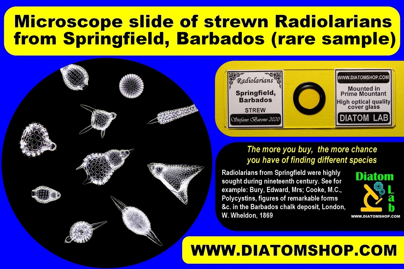
The more you buy, the more chance you have of finding different species!Microscope slide of strewn Radiolarians from Springfield, Barbados, rare sample, mounted in Prime Mountant, excellent high refractive index mountant with refractive index > 1,7. Radiolarians (Polycystine) from Springfield were highly sought during nineteenth century, see for example: 1) Bury, Edward, Mrs; Cooke, M.C., Polycystins, figures of remarkable forms &c. in the Barbados chalk deposit (chiefly collected by Dr. Davy, and noticed in a lecture delivered to the Agricultural Society of Barbados, in July, 1846), London, W. Wheldon, 1869; 2) Association for Advancement of Science, Report of the Seventeenth Meeting of the British Association for the Advancement of Science, held at Oxford in June 1847, London, John Murray, 1848
69.00 €
Add
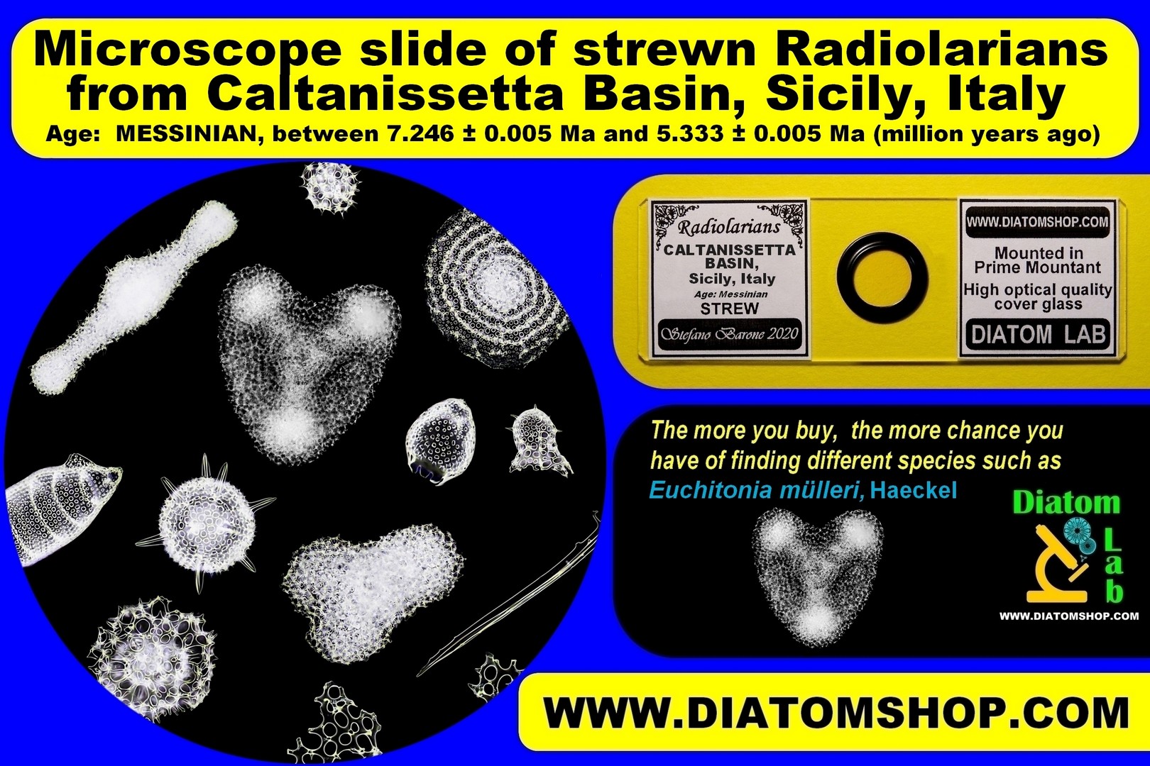
The more you buy, the more chance you have of finding different species!EXTREMELY RARE Microscope slide of strewn Radiolarians from Caltanissetta Basin, Sicily, Italy. From Messinian deposit: 7.246 ± 0.005 - 5.333 ± 0.005 Ma (million years ago). The slide contains several very rare Radiolarian species, including Euchitonia mülleri, Haeckel. Mounted in Prime Mountant, excellent high refractive index mountant with refractive index > 1,7.
59.00 €
Add
The more you buy, the more chance you have of finding different species! Microscope slide of strewn Radiolarians from Mount Hillaby, Barbados,
mounted in Prime Mountant, excellent high refractive index mountant with refractive index > 1,7.
mounted in Prime Mountant, excellent high refractive index mountant with refractive index > 1,7.
99.00 €
Add
The more you buy, the more chance you have of finding different species!Microscope slide of strewn Radiolarians from West Indies - sample collected by Cap. Perry, 1890, cleaned in Nineteenth century: Diatom Lab has decided to avoid any further cleaning in its laboratory to respect the great historical value of this sample, however these Radiolarians are clean with just few impurities. Mounted in Prime Mountant, excellent high refractive index mountant with refractive index > 1,7.
59.00 €
Add

10 selected, micromanipulated Radiolarians for Scanning Electron Microscopy (SEM), micromanipulated and fixed on microscope slide without cover glass and mountant in this case. Radiolarians are fixed by means of the proprietary Diatom Lab's NANO-ADHESIVE, which is invisible to scanning electron microscopes and is therefore used to obtain a clean background for our SEM specimen preparations!
299.00 €
Add
MICROSCOPE SLIDES OF FORAMINIFERA
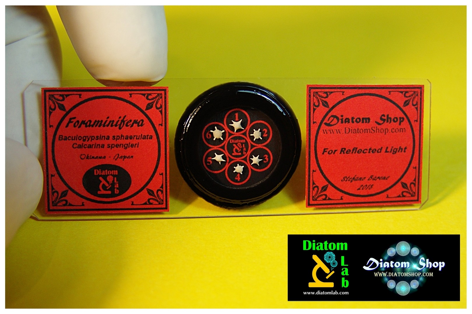
Six selected Foraminifera (Baculogypsina sphaerulata Parker & Jones 1860, Calcarina spengleri Gmelin 1791) from Ryukyu Islands (Japan), dry mounted on microscope slide with protective cover glass. Three colours of labels available (light blue, green, red), write us the colors you like. Only the best, cleanest, unbroken specimens are hand selected! For Stereo microscope or Light microscope in reflected light
69.00 €
Add

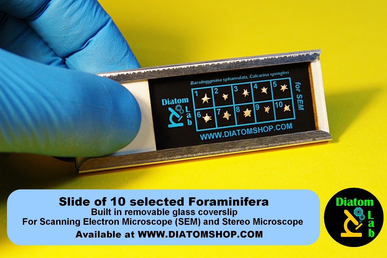
Cardboard slide of 10 selected, micromanipulated Foraminifera (Baculogypsina sphaerulata Parker & Jones 1860, Calcarina spengleri Gmelin 1791) from Ryukyu Islands (Japan), for Stereo microscope. Only the best, cleanest, unbroken specimens are hand selected! Built in removable glass coverslip. Only the best, cleanest, unbroken specimens are hand selected!
149.00 €
Add
MICROSCOPE SLIDES FOR SCANNING ELECTRON MICROSCOPE (SEM) and ATOMIC FORCE MICROSCOPE (AFM).
Venture into the Micro and Nano structures!
10 selected, micromanipulated Diatoms for Scanning Electron Microscope (SEM) and Atomic Force Microscope (AFM), with Species list, micromanipulated and fixed on microscope slide without cover glass and mountant in this case. Diatoms are fixed by means of the proprietary Diatom Lab's NANO-ADHESIVE, which is invisible to scanning electron microscopes and is therefore used to obtain a clean background for our SEM specimen preparations!
299.00 €
Add
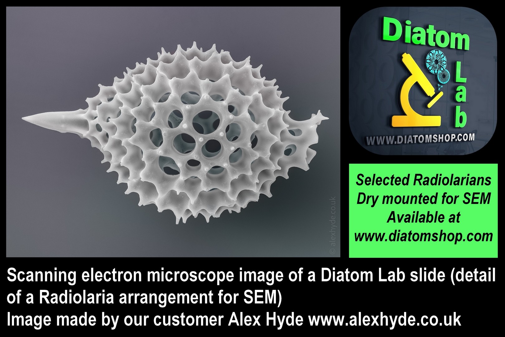
10 selected, micromanipulated Radiolarians for Scanning Electron Microscope (SEM), micromanipulated and fixed on microscope slide without cover glass and mountant in this case. Radiolarians are fixed by means of the proprietary Diatom Lab's NANO-ADHESIVE, which is invisible to scanning electron microscopes and is therefore used to obtain a clean background for our SEM specimen preparations!
299.00 €
Add
Cardboard slide of 10 selected, micromanipulated Foraminifera (Baculogypsina sphaerulata Parker & Jones 1860, Calcarina spengleri Gmelin 1791) from Ryukyu Islands (Japan), for Scanning Electron Microscope (SEM) and Stereo microscope. Only the best, cleanest, unbroken specimens are hand selected! Built in removable glass coverslip. Only the best, cleanest, unbroken specimens are hand selected!
149.00 €
Add
TEST INTEGRATED CIRCUITS for light microscopes in reflected light, stereo microscopes, scanning electron microscopes (SEMs), objectives for macro photography
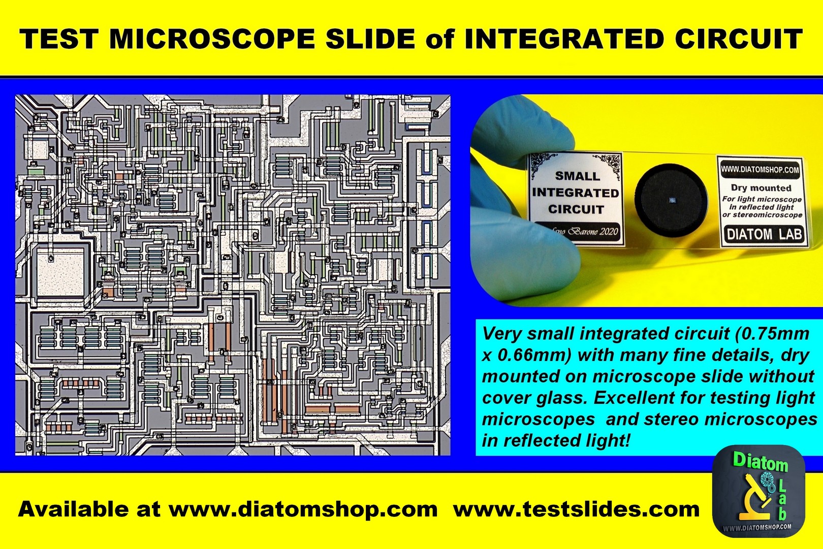
Very small integrated circuit (0.75mm X 0.66mm) with many fine details, dry mounted on microscope slide without cover glass. Excellent for testing light microscopes in reflected light, stereo microscopes, scanning electron microscopes (SEMs), objectives for macro photography! This test integrated circuit has been discussed in the following article: Walker D., Exploring the Diatom Lab prepared slide of an integrated circuit chip, Micscape Magazine, April 2020, Issue 289, ISSN 1365 – 070x: "The slide is impeccably prepared / mounted", "In my view the slide is excellent value for money and maintains the high standards set by the Diatom Lab shop (I have a number of the diatom slides)"
69.00 €
Add
OTHER MICROSCOPE SLIDES
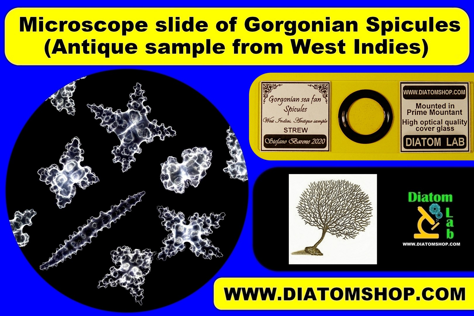
Strew microscope slide of Gorgonian Spicules. Rare antique sample from West Indies. Look at the old papers in the second pictures on the side. Mounted in Prime Mountant, excellent high refractive index mountant with refractive index > 1,7.
49.00 €
Add

Microscope slide of six mixed, micromanipulated Silicoflagellates from Oamaru (NZ) (mounted in Diatom Cubed mountant, refractive index > 1,7). Look at the example on the side.
149.00 €
Add
Microscope slide of six mixed, micromanipulated Silicoflagellates from various locations, arranged in a line or in circle (mounted in Diatom Cubed mountant, refractive index > 1,7).
149.00 €
Add
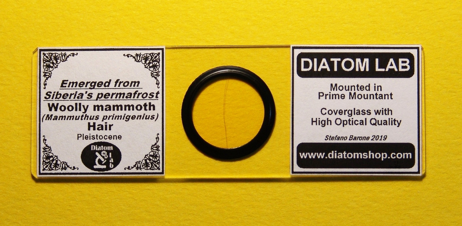
Microscope slide of Wooly Mammoth hair (Mammuthus primigenius), specimen emerged from Siberia's permafrost (Pleistocene). Mounted in Prime Mountant.
59.00 €
Add
DIATOM LAB POSTER
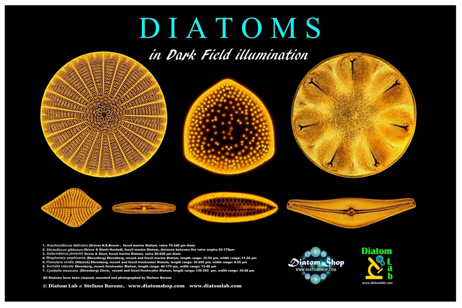
Diatom Lab poster (Diatoms in dark field with scientific names). High resolution microscope image. High quality photographic paper. 12 inch x 8 inch (30,4 cm x 20,3 cm)
49.00 €
Add
CUSTOMIZED ARRANGEMENTS, SERVICES
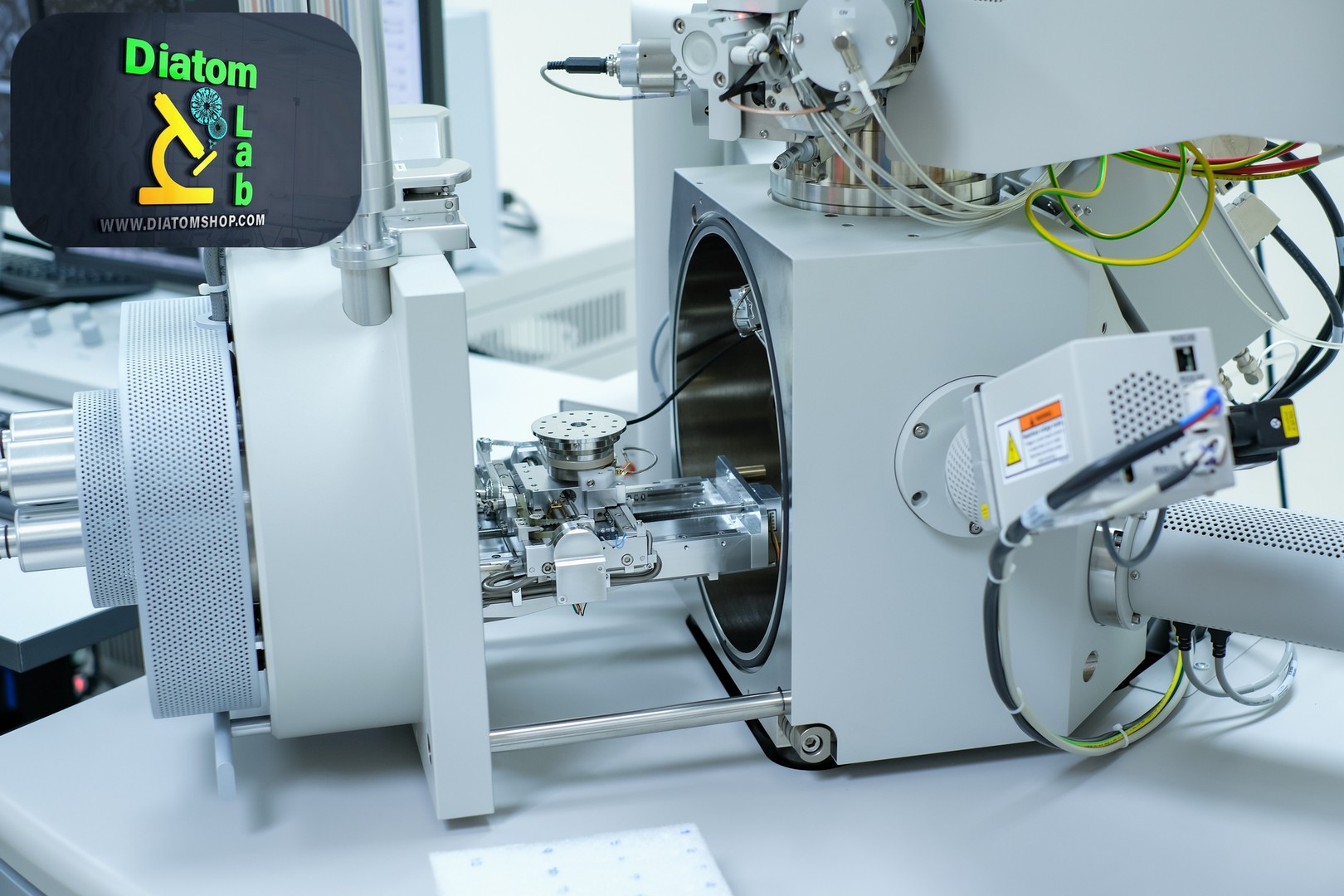
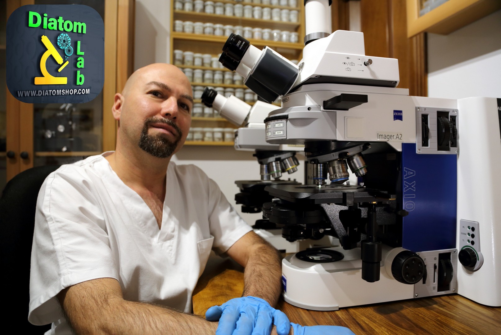
High-resolution microscope images and microscope videos (exclusive or non-exclusive licences): Diatom Lab has won several international scientific photography awards. We provide scientific imaging services using our state of the art Zeiss Axio Imager.A2 and other microscopes in full frame camera format. Various illumination techniques are available. Price: ask for a free quote
This product is unavailable.
0.00 €
New publications, articles for magazines: ask for a free quote
This product is unavailable.
0.00 €
Specifically mounted diatom frustules for photonic researches and applications. In fact Diatoms are photonic crystals (photonics is the skill of manipulating photons): they exhibit sophisticated optical systems. Diatom Lab has collaborated with various institutes by providing specifically mounted diatom frustules for many researches. Price: ask for a free quote
This product is unavailable.
0.00 €
Mounted in the excellent Diatom Cubed mountant (refractive index > 1,7). Price: ask for a free quote
This product is unavailable.
0.00 €
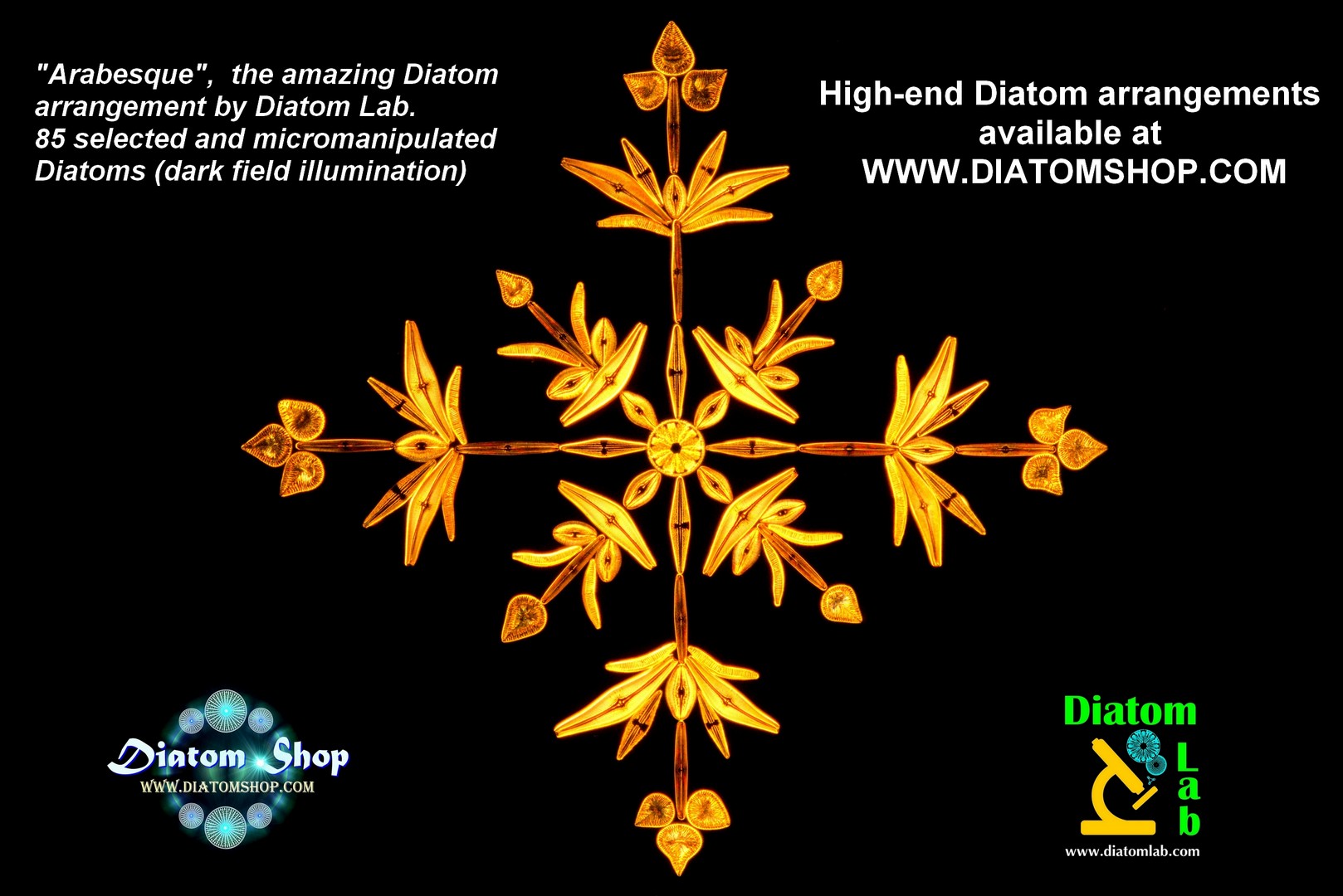
Arabesque, the amazing diatom arrangement (version A), copied exactly from the original: 85 selected Diatoms mounted in the excellent Diatom Cubed mountant (refractive index > 1,7). This arranged diatom slide has been exhibited at Scienza & Natura Expo, 25-26 March 2017 (Novegro, Italy). See photomicrograph in darkfield illumination on the side. Price: ask for a free quote
This product is unavailable.
0.00 €


Please feel free to contact us with questions regarding quotes for customized products and services. We will do our best to answer your request as soon as possible.
"The revelations of the Microscope are perhaps not excelled in importance by those of telescope. While exciting our curiosity, our wonder and admiration, they have proved of infinite service in advancing our knowledge of things around us"(Joseph Leidy)
