THE 10 REASONS DIATOM LAB PREPARATIONS ARE SO INNOVATIVE:
REASON 4: Custom optical quality cover glass is used in all our Diatom preparations. Our coverslips are manufactured to strict tolerances in Germany specifically for Diatom Lab with the following benefits: High transparency, optical homogeneity (freedom from inclusions, bubbles, streaks, etc.), high transmission, excellent flatness, chemical stability, and a refraction index adjusted for microscopy.
REASON 6: As for micromanipulations, only the best, cleanest, unbroken specimens are hand selected, and all slides are quality checked on our in-house Zeiss Axio Imager.A2 research microscope before shipment: Diatom Lab offers perfectly cleaned, selected Diatoms, Radiolarians and other microscopic objects that are micromanipulated paying attention to their ideal positioning to show off the best features.
REASON 8: Diatom Lab's special techniques and Diatom Cubed mountant ensure that Diatoms and Radiolarians will never change their original position during shipping or storage. In other words, you will never see "floating" Diatoms and Radiolarians.
REASON 9: Diatom Lab's unique ringing cements are resistant to the most common immersion oils used in optical microscopy.
Diatom Lab preparations allows you to observe Diatoms and other microscopic objects in Very High Definition! Our prepared microscope slides of Micromanipulated Diatoms and Radiolarians will last for generations, and are produced with modern, state-of-the art techniques and mounting media. We have greatly improved on the "old" processes of mounting and arranging Diatoms and other microscopic specimens, in order to meet the requirements of modern high performance microscopes, for a truly extraordinary visual experience!
Our innovative, state-of-the-art, never published to date, formulas and technologies are the reason for our success among many laboratories and universities in the world. Our materials and methods are protected by trade secret under article 39 of the the TRIPS Agreement.
Diatom Lab premium quality microscope slides are manufactured in our own laboratory under rigorous scientific control! Only the best glasses are used: custom optical quality cover glasses and 26 x 76 mm (1 x 3 inch) fine microscope slides with ground edges. Diatoms and Radiolarians are microscopic, jewel-like products of nature but they must be prepared with the best techniques, tools and materials in order to admire them in all their full glory!
REASON 1: Our Micromanipulated Diatoms and Radiolarians are guaranteed to be fixed directly to the UNDERSIDE of the cover glass (and not, as is common, on the microscope slide) for maximum resolution and contrast! The reason for this: microscope objective performance drops quickly noticeably as the specimen distance from the cover glass increases. Microscope objective lenses are designed to be optimally corrected for objects located immediately below the coverslip! (source: Hiraoka Y., Sedat J. W. and Agard, D. A. (1990) Determination of three-dimensional imaging properties of a light microscope system, Biophysical Journal, 57, 325-333).
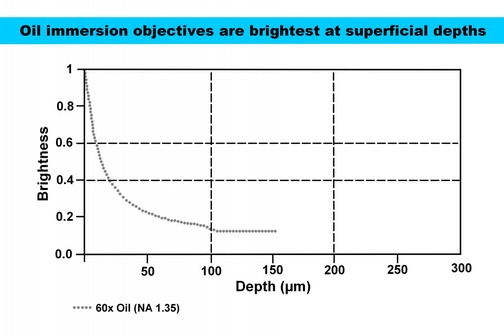
Moreover Oil immersion objectives are brightest at superficial depths, consider the following graph (source: Olympus https://www.olympus-lifescience.com).
Moreover Oil immersion objectives are brightest at superficial depths, consider the following graph (source: Olympus https://www.olympus-lifescience.com).
Furthermore, mounting specimens to the UNDERSIDE of the cover glass allows use of high numerical aperture objective such as oil immersion objectives, even those with very short working distance, such 63x/1,4 or 100x/1,4 apochromats for the ultimate in resolution and contrast!
REASON 2: We use the proprietary DIATOM³ (DIATOM CUBED) high refractive index microfiltered diatom mountant with a refractive index of over 1.7 and excellent chemical stability!
REASON 3: We use the proprietary NANO-ADHESIVE. Diatoms are attached to the underside of the cover glass by means of Diatom Lab's unique NANO-ADHESIVE, which is invisible even in phase contrast, darkfield illumination, differential interference contrast (DIC) and oblique illumination! This ensures all the finest Diatom details such as areolae, poroids, striae and so-on, are always perfectly clean, regardless of the technique used, with the additional benefit that there are no traces, stains or swipe-marks caused by fixatives typically seen on some older preparations. Futhermore, our NANO-ADHESIVE is also invisible to scanning electron microscopes (SEMs), and is therefore used to obtain a clean background for our SEM specimen preparations. See the test image below, performed by Purdue University Fort Wayne on Diatom Lab's preparations that contain Diatoms fixed on glass using Diatom Lab's NANO-ADHESIVE (without mountant and cover glass to permit SEM imaging):
[image:image-1]
REASON 4: Custom optical quality cover glass is used in all our Diatom preparations. Our coverslips are manufactured to strict tolerances in Germany specifically for Diatom Lab with the following benefits: High transparency, optical homogeneity (freedom from inclusions, bubbles, streaks, etc.), high transmission, excellent flatness, chemical stability, and a refraction index adjusted for microscopy.
REASON 5: Diatom Lab's microscope slides are prepared taking into account the cover glass thickness suitable for each preparation, in fact: using the correct coverslip thickness can greatly increase your ability to get the most information out of your sample using an optical microscope (source: Douglas W. Cromey, M.S. (2017) The importance of #1.5 thickness coverslips for Microscopy, UA Microscopy Alliance, The University of Arizona, Tucson, AZ)
REASON 6: As for micromanipulations, only the best, cleanest, unbroken specimens are hand selected, and all slides are quality checked on our in-house Zeiss Axio Imager.A2 research microscope before shipment: Diatom Lab offers perfectly cleaned, selected Diatoms, Radiolarians and other microscopic objects that are micromanipulated paying attention to their ideal positioning to show off the best features.
All recent and fossil Diatoms and Radiolarians are cleaned in our laboratory using innovative, and professional techniques.
REASON 7: Diatom lab's Diatoms and Radiolarians can be observed and admired even with a modestly priced microscope (contrast enhancement techniques such as phase contrast, darkfield illumination and differential interference contrast are not strictly necessary) thanks to Diatom Lab's strict quality procedures.
REASON 8: Diatom Lab's special techniques and Diatom Cubed mountant ensure that Diatoms and Radiolarians will never change their original position during shipping or storage. In other words, you will never see "floating" Diatoms and Radiolarians.
REASON 9: Diatom Lab's unique ringing cements are resistant to the most common immersion oils used in optical microscopy.
REASON 10: Diatom Lab's Preparations are guaranteed free of bubbles and impurities.